
Search Count: 257
 |
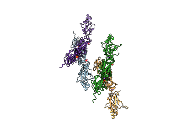
Icosahedral Symmetric Structure Of An Expansion Intermediate Of Turnip Crinkle Virus (Asymmetric Trimer Unit)
Organism: Turnip crinkle virus
Method: ELECTRON MICROSCOPY Release Date: 2025-11-12 Classification: VIRUS |
 |
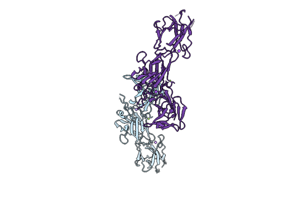
Symmetry Relaxed Asymmetric Structure Of An Expansion Intermediate Of Turnip Crinkle Virus
Organism: Turnip crinkle virus
Method: ELECTRON MICROSCOPY Release Date: 2025-11-12 Classification: VIRAL PROTEIN |
 |
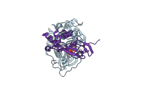
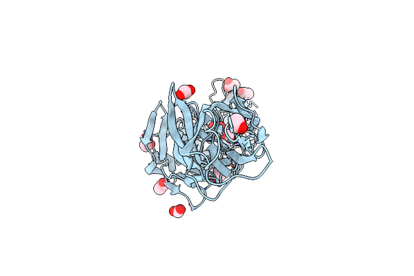
Organism: Thermobifida fusca yx
Method: X-RAY DIFFRACTION Release Date: 2025-08-06 Classification: HYDROLASE |
 |
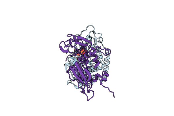
Organism: Thermobifida fusca yx
Method: X-RAY DIFFRACTION Release Date: 2025-07-23 Classification: HYDROLASE Ligands: CL, NA, GOL |
 |
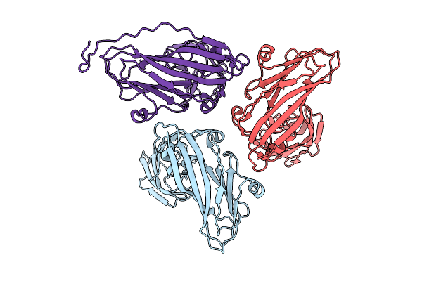
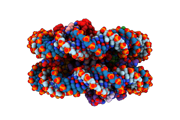
Crystal Structure Of C-Terminal Rigid Fragment Containing Middle And C-Terminal Domains Of The Basal Pilin Ebpb From Enterococcus Faecalis.
Organism: Enterococcus faecalis og1rf
Method: X-RAY DIFFRACTION Release Date: 2025-07-16 Classification: CELL ADHESION |
 |
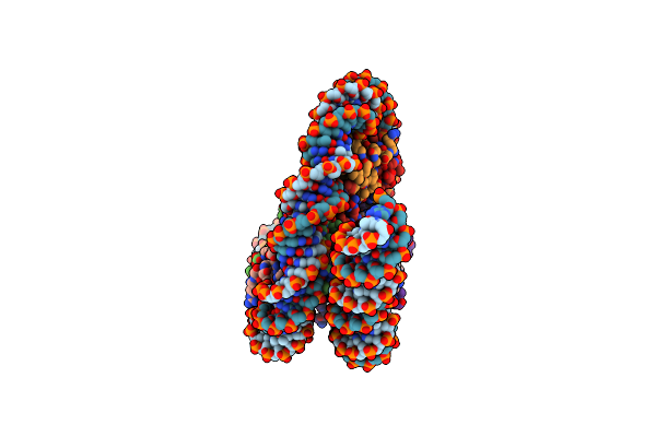
Crystal Structure Of N-Terminal Flexible Domain Of The Shaft Pilin Ebpc From Enterococcus Faecalis.
Organism: Enterococcus faecalis og1rf
Method: X-RAY DIFFRACTION Release Date: 2025-07-16 Classification: CELL ADHESION |
 |
Crystal Structure Of N-Terminal Flexible Domain Of The Shaft Pilin Ebpc From Enterococcus Faecalis.
Organism: Enterococcus faecalis og1rf
Method: X-RAY DIFFRACTION Release Date: 2025-07-16 Classification: CELL ADHESION |
 |
Organism: Enterococcus faecalis og1rf
Method: X-RAY DIFFRACTION Release Date: 2025-07-16 Classification: CELL ADHESION |
 |
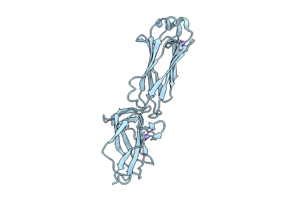
Crystal Structure Of C-Terminal Stable Fragment Of The Shaft Pilin Ebpc From Enterococcus Faecalis
Organism: Enterococcus faecalis og1rf
Method: X-RAY DIFFRACTION Release Date: 2025-07-16 Classification: CELL ADHESION Ligands: MG, NA |
 |
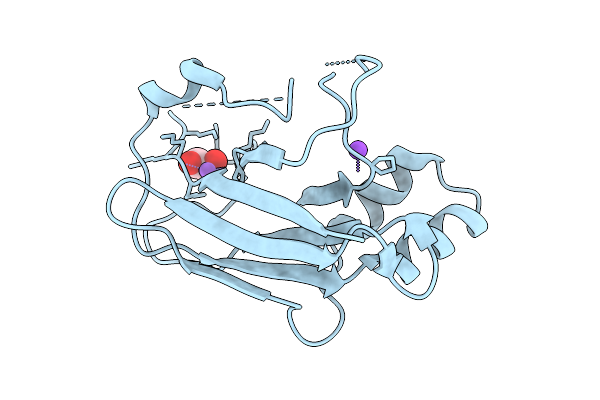
Crystal Structure Of The Basal Pilin Ebpb From Enterococcus Faecalis With A Partially Disordered N-Terminal Domain.
Organism: Enterococcus faecalis og1rf
Method: X-RAY DIFFRACTION Release Date: 2025-07-16 Classification: CELL ADHESION |
 |
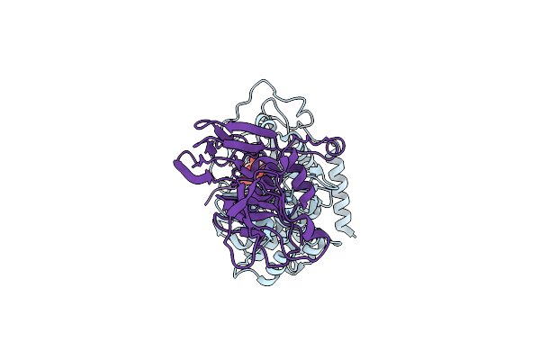
Organism: Thermobifida fusca yx
Method: X-RAY DIFFRACTION Release Date: 2025-07-09 Classification: HYDROLASE Ligands: EDO, CL, NA |
 |
Structure Of A Native Drosophila Melanogaster Pol Ii Elongation Complex Without Rpb4/Rpb7 Stalk
Organism: Drosophila melanogaster
Method: ELECTRON MICROSCOPY Resolution:3.37 Å Release Date: 2025-05-14 Classification: TRANSCRIPTION Ligands: ZN |
 |
Organism: Streptococcus sanguinis sk36
Method: X-RAY DIFFRACTION Resolution:2.00 Å Release Date: 2025-05-07 Classification: STRUCTURAL PROTEIN |
 |
Organism: Homo sapiens, Bos taurus
Method: ELECTRON MICROSCOPY Release Date: 2025-04-23 Classification: SIGNALING PROTEIN Ligands: ATP |
 |
Organism: Homo sapiens, Bos taurus
Method: ELECTRON MICROSCOPY Release Date: 2025-04-23 Classification: SIGNALING PROTEIN Ligands: ATP |
 |
Organism: Homo sapiens, Bos taurus
Method: ELECTRON MICROSCOPY Release Date: 2025-04-23 Classification: SIGNALING PROTEIN Ligands: ATP |
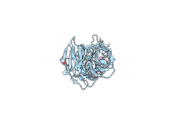 |
Crystal Structure Of Major Pilin Pilb From Streptococcus Sanguinis - Semet Derivative
Organism: Streptococcus sanguinis sk36
Method: X-RAY DIFFRACTION Resolution:2.40 Å Release Date: 2025-03-26 Classification: CELL ADHESION Ligands: EDO, ACT, NA |
 |
Organism: Streptococcus sanguinis sk36
Method: X-RAY DIFFRACTION Resolution:1.88 Å Release Date: 2025-03-26 Classification: CELL ADHESION Ligands: FMT, ACY, EDO, ACT, NA |
 |
Organism: Drosophila melanogaster
Method: ELECTRON MICROSCOPY Release Date: 2025-02-19 Classification: GENE REGULATION |
 |
Organism: Drosophila melanogaster
Method: ELECTRON MICROSCOPY Release Date: 2025-02-19 Classification: GENE REGULATION |