
Search Count: 81
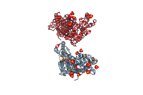 |
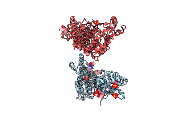
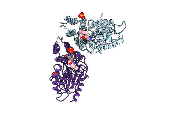
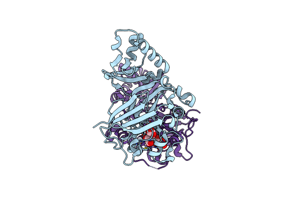
Mycobacterium Tuberculosis Pks13 Acyltransferase Serine Converted To Beta-Lactam Form By Cec215 Via Sufex Reaction
Organism: Mycobacterium tuberculosis h37rv
Method: X-RAY DIFFRACTION Resolution:2.22 Å Release Date: 2025-05-21 Classification: ANTIBIOTIC Ligands: SO4, CL, PG4, EDO, DMS, GOL, PEG |
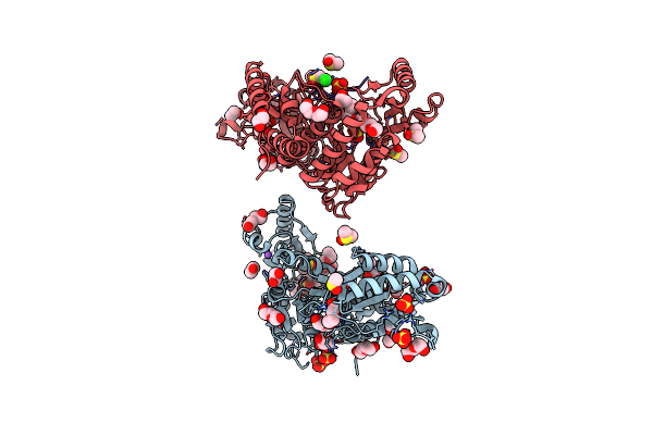 |
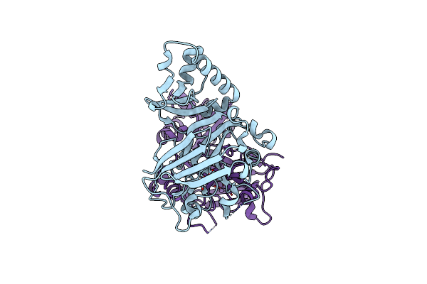
Organism: Mycobacterium tuberculosis (strain atcc 25618 / h37rv)
Method: X-RAY DIFFRACTION Resolution:2.05 Å Release Date: 2025-05-21 Classification: ANTIBIOTIC Ligands: DMS, SO4, PE5, EDO, PEG, GOL, CL, NA, P33 |
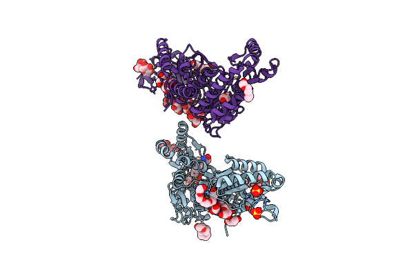 |
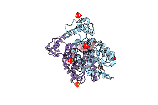
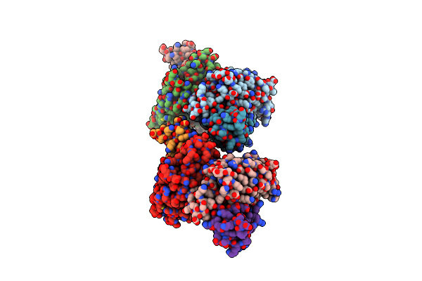
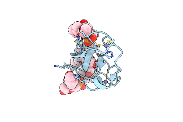
M. Tuberculosis Pks13 Acyltransferase (At) Domain In Complex With Sufex Inhibitor Cec215
Organism: Mycobacterium tuberculosis
Method: X-RAY DIFFRACTION Resolution:1.87 Å Release Date: 2025-05-07 Classification: TRANSFERASE/TRANSFERASE INHIBITOR Ligands: 1PE, SO4, A1ATV |
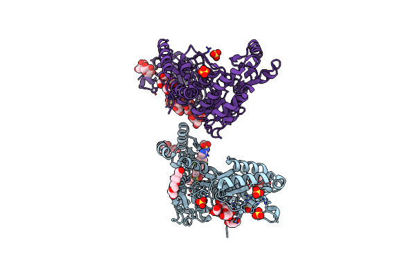 |
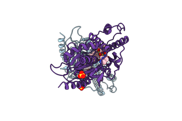
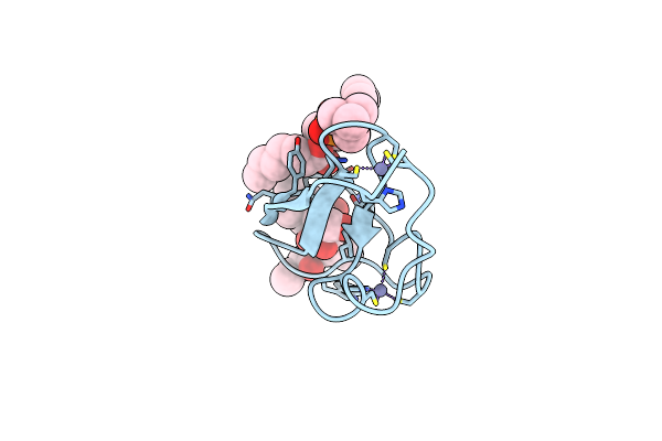
M. Tuberculosis Pks13 Acyltransferase (At) Domain In Complex With Sufex Inhibitor Cmx410
Organism: Mycobacterium tuberculosis (strain atcc 25618 / h37rv)
Method: X-RAY DIFFRACTION Resolution:2.57 Å Release Date: 2025-05-07 Classification: TRANSFERASE/TRANSFERSE INHIBITOR Ligands: 1PE, SO4, A1ATW |
 |
Organism: Mycobacterium tuberculosis
Method: X-RAY DIFFRACTION Resolution:2.18 Å Release Date: 2025-05-07 Classification: TRANSFERASE Ligands: 1PE, SO4 |
 |
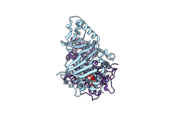
M. Tuberculosis Pks13 Acyltransferase (At) Domain In Complex With Sufex Inhibitor Cmx410 - Reaction Product
Organism: Mycobacterium tuberculosis (strain atcc 25618 / h37rv)
Method: X-RAY DIFFRACTION Resolution:2.02 Å Release Date: 2025-05-07 Classification: TRANSFERASE Ligands: SO4, 1PE, A1AVL |
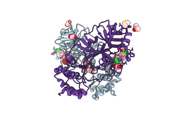 |
Organism: Severe acute respiratory syndrome coronavirus 2
Method: X-RAY DIFFRACTION Resolution:1.76 Å Release Date: 2024-11-20 Classification: HYDROLASE/INHIBITOR Ligands: DMS, EDO, SO4, A1A21, A1AFD, GOL |
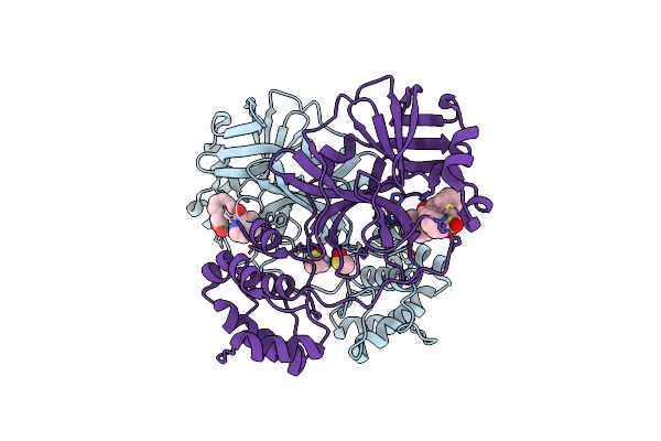 |
Organism: Severe acute respiratory syndrome coronavirus 2
Method: X-RAY DIFFRACTION Resolution:2.05 Å Release Date: 2024-11-20 Classification: HYDROLASE/INHIBITOR Ligands: A1AFE, DMS |
 |
Crystal Structure Of Mtb Pks13 Thioesterase Domain In Complex With Inhibitor X20419
Organism: Mycobacterium tuberculosis
Method: X-RAY DIFFRACTION Resolution:2.20 Å Release Date: 2024-04-24 Classification: BIOSYNTHETIC PROTEIN Ligands: JR0, SO4 |
 |
Crystal Structure Of Mtb Pks13 Thioesterase Domain In Complex With Inhibitor X20403
Organism: Mycobacterium tuberculosis h37rv
Method: X-RAY DIFFRACTION Resolution:2.00 Å Release Date: 2024-04-24 Classification: BIOSYNTHETIC PROTEIN Ligands: JS9, SO4 |
 |
Crystal Structure Of Mtb Pks13 Thioesterase Domain In Complex With Inhibitor X20404
Organism: Mycobacterium tuberculosis
Method: X-RAY DIFFRACTION Resolution:2.10 Å Release Date: 2024-04-24 Classification: BIOSYNTHETIC PROTEIN Ligands: K6C, SO4 |
 |
Crystal Structure Of Mtb Pks13 Thioesterase Domain In Complex With Inhibitor X20348
Organism: Mycobacterium tuberculosis
Method: X-RAY DIFFRACTION Resolution:2.35 Å Release Date: 2024-04-24 Classification: BIOSYNTHETIC PROTEIN Ligands: QU0, SO4 |
 |
Organism: Bacillus subtilis
Method: X-RAY DIFFRACTION Resolution:2.40 Å Release Date: 2023-11-01 Classification: HYDROLASE Ligands: OXL, MN |
 |
Crystal Structure Of Yisk From Bacillus Subtilis In Complex With Reaction Product 4-Hydroxy-2-Oxoglutaric Acid
Organism: Bacillus subtilis
Method: X-RAY DIFFRACTION Resolution:1.93 Å Release Date: 2023-11-01 Classification: HYDROLASE Ligands: IO9, MN |
 |
Organism: Bacillus subtilis
Method: X-RAY DIFFRACTION Resolution:2.26 Å Release Date: 2023-11-01 Classification: HYDROLASE Ligands: MN |
 |
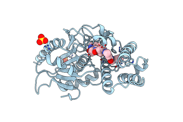
Mycobacterium Tuberculosis 3-Hydroxyl-Acp Dehydratase Hadab In Complex With 1,3-Diarylpyrazolyl-Acylsulfonamide Inhibitor
Organism: Mycobacterium tuberculosis
Method: X-RAY DIFFRACTION Resolution:2.40 Å Release Date: 2022-11-16 Classification: LYASE/INHIBITOR Ligands: CI7, EDO, PEG, GOL |
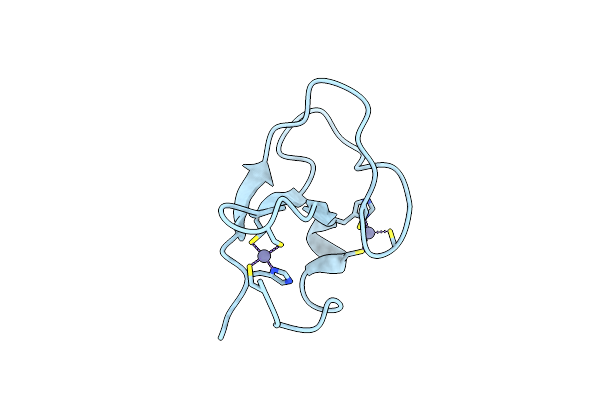 |
Organism: Rattus norvegicus
Method: X-RAY DIFFRACTION Resolution:1.39 Å Release Date: 2022-05-04 Classification: LIPID BINDING PROTEIN Ligands: ZN |
 |
C1B Domain Of Protein Kinase C In Complex With Phorbol Ester And Phosphatidylcholine
Organism: Rattus norvegicus
Method: X-RAY DIFFRACTION Resolution:1.57 Å Release Date: 2022-05-04 Classification: LIPID BINDING PROTEIN Ligands: XP5, ZN, WTS |
 |
C1B Domain Of Protein Kinase C In Complex With Ingenol-3-Angelate And Phosphocholine
Organism: Rattus norvegicus
Method: X-RAY DIFFRACTION Resolution:1.80 Å Release Date: 2022-05-04 Classification: LIPID BINDING PROTEIN Ligands: ZN, WUD, XP5 |
 |
C1B Domain Of Protein Kinase C In Complex With Diacylglycerol And Dodecyl 2-(Trimethylammonio)Ethyl Phosphate
Organism: Rattus norvegicus
Method: X-RAY DIFFRACTION Resolution:1.75 Å Release Date: 2022-05-04 Classification: LIPID BINDING PROTEIN Ligands: ZN, DGA, EOH, DPV |