
Search Count: 35
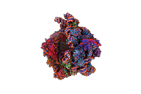 |
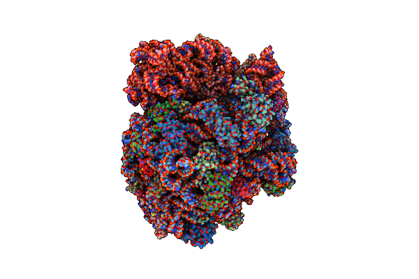
Organism: Escherichia coli, Vibrio alginolyticus
Method: ELECTRON MICROSCOPY Release Date: 2022-06-22 Classification: RIBOSOME Ligands: MG, K |
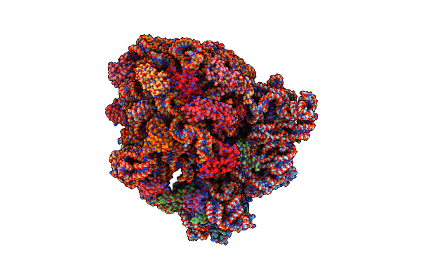 |
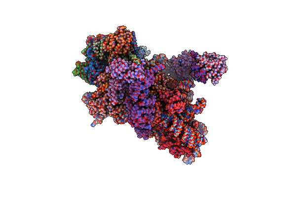
Organism: Escherichia coli, Vibrio alginolyticus, Escherichia coli k-12, Escherichia coli umea 3162-1
Method: ELECTRON MICROSCOPY Release Date: 2022-04-27 Classification: RIBOSOME Ligands: MG, K |
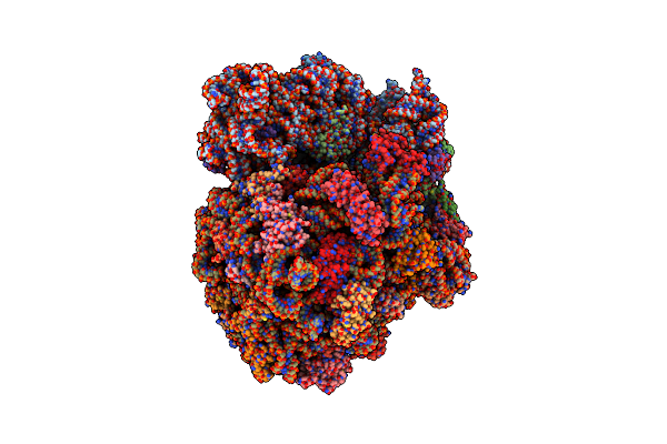 |
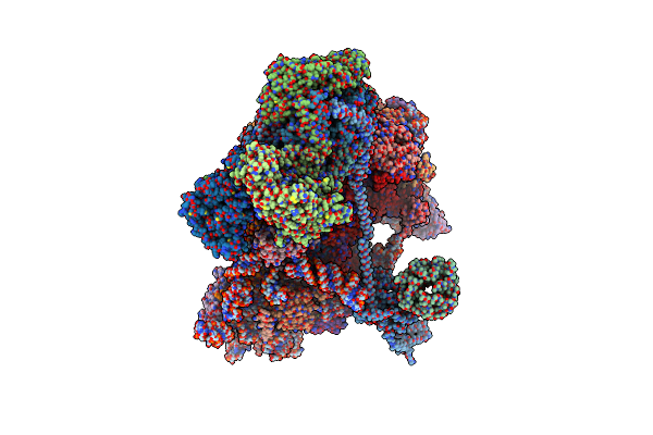
Organism: Vibrio alginolyticus, Escherichia coli k-12
Method: ELECTRON MICROSCOPY Release Date: 2022-03-16 Classification: RIBOSOME Ligands: MG, K |
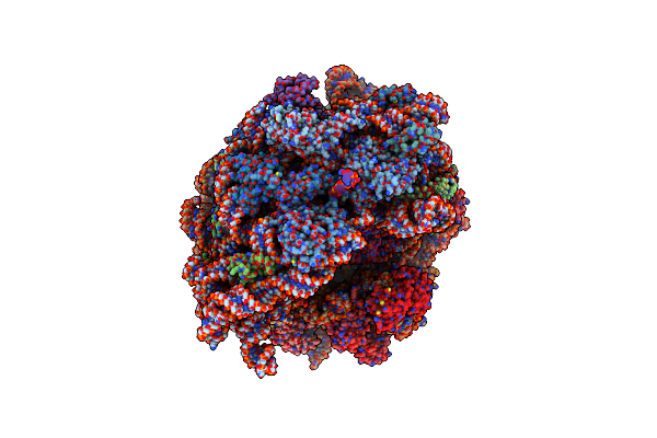 |
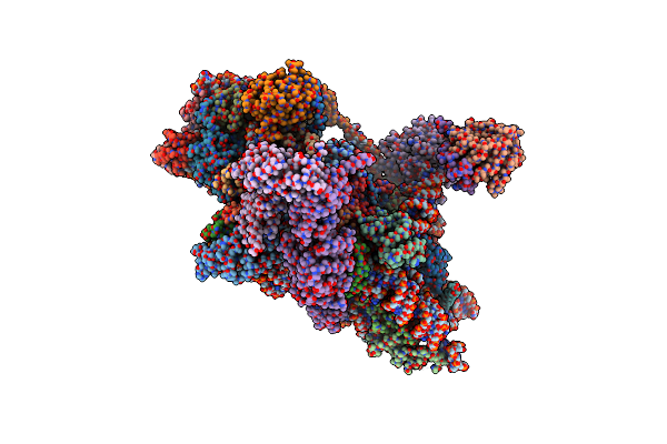
Organism: Escherichia coli k-12
Method: ELECTRON MICROSCOPY Release Date: 2022-03-16 Classification: RIBOSOME Ligands: MG, K |
 |
Organism: Bacillus subtilis
Method: ELECTRON MICROSCOPY Release Date: 2022-03-16 Classification: RIBOSOME |
 |
Organism: Bacillus subtilis
Method: ELECTRON MICROSCOPY Release Date: 2022-03-16 Classification: RIBOSOME |
 |
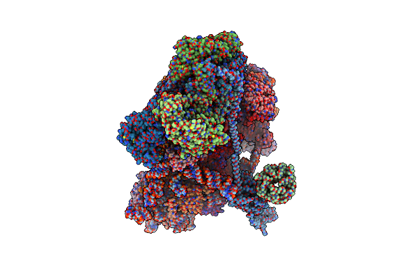
Cryo-Em Structure Of A Bacillus Subtilis Mifm-Stalled Ribosome-Nascent Chain Complex With (P)Ppgpp-Srp Bound
|
 |
Organism: Bacillus subtilis (strain 168)
Method: X-RAY DIFFRACTION Resolution:2.51 Å Release Date: 2022-02-02 Classification: TRANSLATION Ligands: G4P, MG |
 |
Organism: Escherichia coli
Method: X-RAY DIFFRACTION Resolution:2.80 Å Release Date: 2022-02-02 Classification: TRANSLATION Ligands: G4P, MG |
 |
Organism: Escherichia coli dh5[alpha]
Method: X-RAY DIFFRACTION Resolution:2.40 Å Release Date: 2022-02-02 Classification: TRANSLATION Ligands: 0O2 |
 |
Organism: Escherichia coli (strain k12)
Method: X-RAY DIFFRACTION Resolution:2.49 Å Release Date: 2022-02-02 Classification: TRANSLATION Ligands: 0O2 |
 |
Organism: Saccharomyces cerevisiae (strain atcc 204508 / s288c)
Method: ELECTRON MICROSCOPY Release Date: 2020-10-28 Classification: RIBOSOME Ligands: MG, ZN, ADP, ATP, SF4 |
 |
Organism: Homo sapiens
Method: ELECTRON MICROSCOPY Release Date: 2020-10-14 Classification: RIBOSOME Ligands: ZN, SF4, ADP, MG, ATP, MET, GTP |
 |
Organism: Saccharomyces cerevisiae s288c, Saccharomyces cerevisiae (strain atcc 204508 / s288c)
Method: ELECTRON MICROSCOPY Release Date: 2020-10-14 Classification: RIBOSOME Ligands: MG, ZN, SF4, ADP |
 |
Organism: Saccharomyces cerevisiae (strain atcc 204508 / s288c), Saccharomyces cerevisiae s288c
Method: ELECTRON MICROSCOPY Release Date: 2020-10-07 Classification: RIBOSOME Ligands: ZN, ADP, MG, ATP, SF4 |
 |
Organism: Homo sapiens
Method: ELECTRON MICROSCOPY Release Date: 2020-10-07 Classification: RIBOSOME Ligands: ZN, SF4, ADP, MG, ATP |
 |
Organism: Homo sapiens, Severe acute respiratory syndrome coronavirus 2
Method: ELECTRON MICROSCOPY Release Date: 2020-08-19 Classification: VIRAL PROTEIN Ligands: MG, ZN |
 |
Organism: Homo sapiens, Severe acute respiratory syndrome coronavirus 2
Method: ELECTRON MICROSCOPY Release Date: 2020-08-19 Classification: VIRAL PROTEIN Ligands: MG, ZN |
 |
Organism: Homo sapiens, Severe acute respiratory syndrome coronavirus 2
Method: ELECTRON MICROSCOPY Release Date: 2020-08-19 Classification: VIRAL PROTEIN Ligands: ZN |
 |
Organism: Homo sapiens, Severe acute respiratory syndrome coronavirus 2
Method: ELECTRON MICROSCOPY Release Date: 2020-08-12 Classification: VIRAL PROTEIN Ligands: MG, ZN, SF4, ADP |