
Search Count: 25
 |
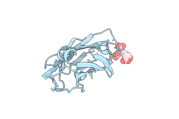
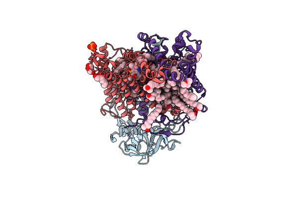
Fimh In Complex With Alpha1,6 Core-Fucosylated Oligomannose-3, Crystallized In The Trigonal Space Group
Organism: Escherichia coli k-12
Method: X-RAY DIFFRACTION Resolution:1.45 Å Release Date: 2023-04-12 Classification: CELL ADHESION Ligands: NI, SO4 |
 |
Organism: Escherichia coli k-12
Method: X-RAY DIFFRACTION Resolution:3.19 Å Release Date: 2023-04-12 Classification: CELL ADHESION Ligands: NI, SO4 |
 |
Organism: Escherichia coli bl21(de3)
Method: X-RAY DIFFRACTION Resolution:3.00 Å Release Date: 2023-02-01 Classification: CELL ADHESION Ligands: NI, SO4 |
 |
Fimh In Complex With Alpha1,6 Core-Fucosylated Oligomannose-3, Crystallized In The Trigonal Space Group
Organism: Escherichia coli (strain k12)
Method: X-RAY DIFFRACTION Resolution:1.40 Å Release Date: 2022-07-20 Classification: CELL ADHESION Ligands: NI, SO4 |
 |
Two Copies Of The Catalytic Domain Of Nana Sialidase From Streptococcus Pneumoniae Juxtaposed In The P212121 Space Group, In Complex With Dana
Organism: Streptococcus pneumoniae
Method: X-RAY DIFFRACTION Resolution:2.70 Å Release Date: 2020-11-18 Classification: STRUCTURAL PROTEIN Ligands: DAN |
 |
Two Copies Of The Catalytic Domain Of Nana Sialidase From Streptococcus Pneumoniae Juxtaposed In The P212121 Space Group, In Complex With Dana Derivatized With A Peg Linker On The Glycerol Group.
Organism: Streptococcus pneumoniae
Method: X-RAY DIFFRACTION Resolution:1.94 Å Release Date: 2020-11-18 Classification: STRUCTURAL PROTEIN Ligands: R7H, SO4, CL, CA |
 |
Co-Crystals In The P212121 Space Group, Of A Beta-Cyclodextrin Spacered By Triazole Heptyl From Alpha-D-Mannose, With Fimh Lectin At 2.00 A Resolution.
Organism: Escherichia coli
Method: X-RAY DIFFRACTION Resolution:1.96 Å Release Date: 2020-05-06 Classification: CELL ADHESION Ligands: TA5, HP6, MAN |
 |
Breaking Down The Wall: Mutation Of The Tyrosine Gate Of The Universal Escherichia Coli Fimbrial Adhesin Fimh
Organism: Escherichia coli
Method: X-RAY DIFFRACTION Resolution:1.90 Å Release Date: 2017-01-18 Classification: CELL ADHESION Ligands: EDT |
 |
Breaking Down The Wall: Mutation Of The Tyrosine Gate Of The Universal Escherichia Coli Fimbrial Adhesin Fimh
Organism: Escherichia coli
Method: X-RAY DIFFRACTION Resolution:1.42 Å Release Date: 2016-11-30 Classification: CELL ADHESION Ligands: KGM, NA |
 |
Breaking Down The Wall: Mutation Of The Tyrosine Gate Of The Universal Escherichia Coli Fimbrial Adhesin Fimh
Organism: Escherichia coli
Method: X-RAY DIFFRACTION Resolution:2.13 Å Release Date: 2016-11-30 Classification: CELL ADHESION Ligands: 3X8 |
 |
Crystal Structure Of Fimh Lectin Domain With The Tyr48Ala Mutation, In Complex With Heptyl Alpha-D-Mannopyrannoside
Organism: Escherichia coli
Method: X-RAY DIFFRACTION Resolution:2.84 Å Release Date: 2014-10-29 Classification: CELL ADHESION Ligands: KGM |
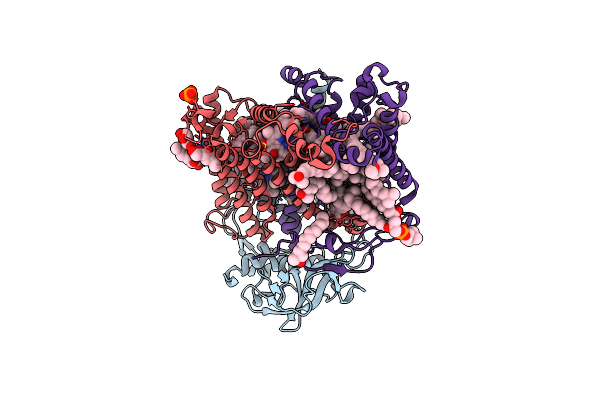 |
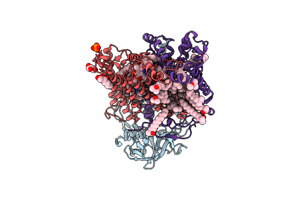
X-Ray High Resolution Structure Of The Photosynthetic Reaction Center From Rb. Sphaeroides At Ph 8 In The Neutral State
Organism: Rhodobacter sphaeroides
Method: X-RAY DIFFRACTION Resolution:1.87 Å Release Date: 2007-07-03 Classification: ELECTRON TRANSPORT Ligands: GOL, BCL, LDA, BPH, U10, PO4, HTO, FE, SPO, CDL, PC1, GGD |
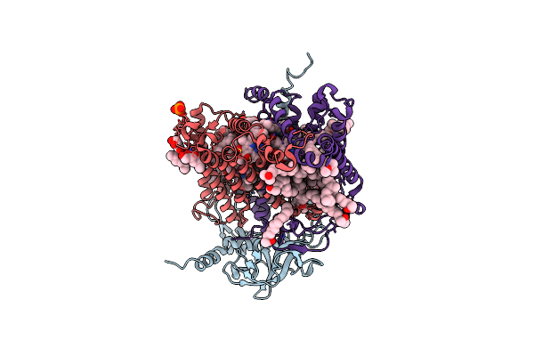 |
X-Ray High Resolution Structure Of The Photosynthetic Reaction Center From Rb. Sphaeroides At Ph 8 In The Charge-Separated State
Organism: Rhodobacter sphaeroides
Method: X-RAY DIFFRACTION Resolution:2.07 Å Release Date: 2007-07-03 Classification: ELECTRON TRANSPORT Ligands: GOL, BCL, LDA, BPH, U10, PO4, HTO, FE, SPO, CDL |
 |
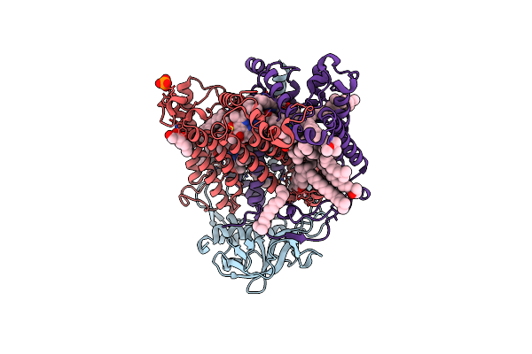
X-Ray High Resolution Structure Of The Photosynthetic Reaction Center From Rb. Sphaeroides At Ph 6.5 In The Charge-Separated State
Organism: Rhodobacter sphaeroides
Method: X-RAY DIFFRACTION Resolution:2.90 Å Release Date: 2007-07-03 Classification: PHOTOSYNTHESIS Ligands: GOL, LDA, BCL, BPH, UQ2, PO4, FE, U10, SPO |
 |
X-Ray High Resolution Structure Of The Photosynthetic Reaction Center From Rb. Sphaeroides At Ph 6.5 In The Charge-Separated State 2Nd Dataset
Organism: Rhodobacter sphaeroides
Method: X-RAY DIFFRACTION Resolution:2.50 Å Release Date: 2007-07-03 Classification: PHOTOSYNTHESIS Ligands: GOL, BCL, BPH, UQ2, PO4, HTO, LDA, FE, U10, SPO |
 |
X-Ray High Resolution Structure Of The Photosynthetic Reaction Center From Rb. Sphaeroides At Ph 6.5 In The Neutral State, 2Nd Dataset
Organism: Rhodobacter sphaeroides
Method: X-RAY DIFFRACTION Resolution:2.04 Å Release Date: 2007-07-03 Classification: PHOTOSYNTHESIS Ligands: GOL, BCL, LDA, BPH, UQ2, PO4, HTO, FE, U10, SPO, CDL |
 |
X-Ray High Resolution Structure Of The Photosynthetic Reaction Center From Rb. Sphaeroides At Ph 6.5 In The Charge-Separated State, 3Rd Dataset
Organism: Rhodobacter sphaeroides
Method: X-RAY DIFFRACTION Resolution:2.13 Å Release Date: 2007-07-03 Classification: PHOTOSYNTHESIS Ligands: GOL, BCL, LDA, BPH, UQ2, PO4, HTO, FE, U10, SPO |
 |
X-Ray High Resolution Structure Of The Photosynthetic Reaction Center From Rb. Sphaeroides At Ph 6.5 In The Neutral State
Organism: Rhodobacter sphaeroides
Method: X-RAY DIFFRACTION Resolution:2.05 Å Release Date: 2007-07-03 Classification: PHOTOSYNTHESIS Ligands: GOL, BCL, LDA, BPH, UQ2, PO4, HTO, FE, U10, SPO, CDL |
 |
X-Ray High Resolution Structure Of The Photosynthetic Reaction Center From Rb. Sphaeroides At Ph 9 In The Neutral State
Organism: Rhodobacter sphaeroides
Method: X-RAY DIFFRACTION Resolution:2.50 Å Release Date: 2007-07-03 Classification: PHOTOSYNTHESIS Ligands: GOL, BCL, LDA, BPH, UQ2, PO4, HTO, FE, U10, SPO, CDL |
 |
X-Ray High Resolution Structure Of The Photosynthetic Reaction Center From Rb. Sphaeroides At Ph 9 In The Charge-Separated State, 2Nd Dataset
Organism: Rhodobacter sphaeroides
Method: X-RAY DIFFRACTION Resolution:2.51 Å Release Date: 2007-07-03 Classification: PHOTOSYNTHESIS Ligands: GOL, BCL, LDA, BPH, UQ2, PO4, HTO, FE, U10, SPO, CDL |