
Search Count: 23
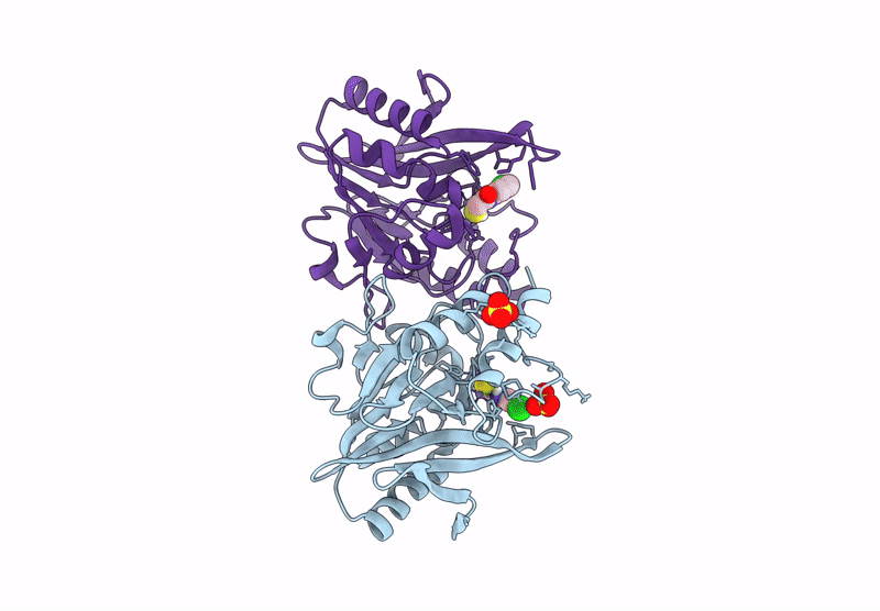 |
Organism: Klebsiella pneumoniae
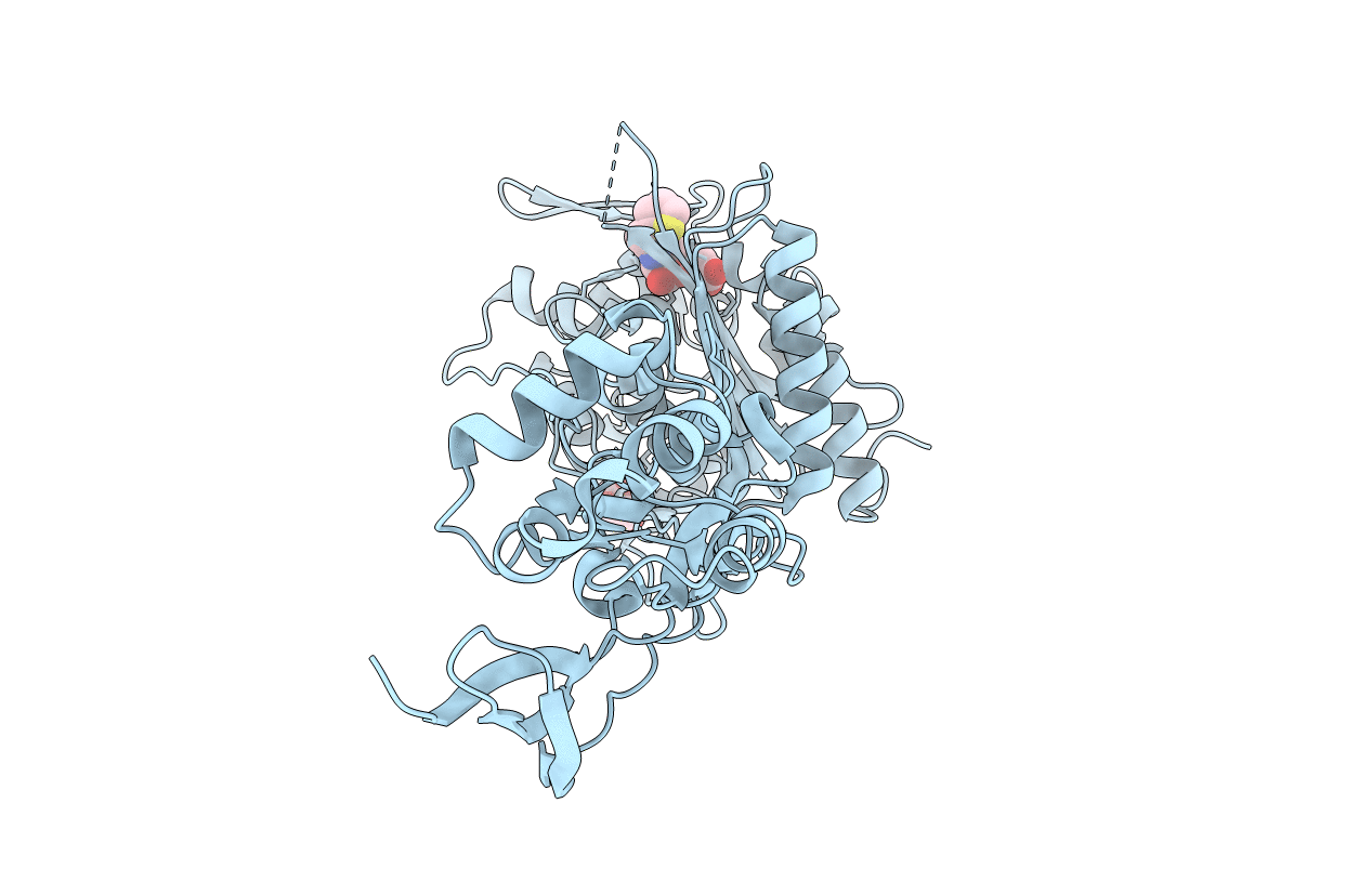
Method: X-RAY DIFFRACTION Release Date: 2025-07-09 Classification: ANTIMICROBIAL PROTEIN Ligands: ZN, SO4, A1JCJ, CL |
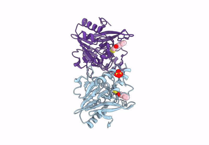 |
Organism: Klebsiella pneumoniae
Method: X-RAY DIFFRACTION Release Date: 2025-07-09 Classification: ANTIMICROBIAL PROTEIN Ligands: ZN, SO4, A1JCK |
 |
Structure Of P. Aeruginosa Pbp3 In Complex With A Phenyl Boronic Acid (Compound 1)
Organism: Pseudomonas aeruginosa pao1
Method: X-RAY DIFFRACTION Resolution:1.58 Å Release Date: 2021-08-11 Classification: HYDROLASE Ligands: GOL, RY2, DMS |
 |
Structure Of P. Aeruginosa Pbp3 In Complex With An Aryl Boronic Acid (Compound 2)
Organism: Pseudomonas aeruginosa pao1
Method: X-RAY DIFFRACTION Resolution:1.59 Å Release Date: 2021-08-11 Classification: HYDROLASE Ligands: RXW, DMS, GOL |
 |
Structure Of P. Aeruginosa Pbp3 In Complex With A Benzoxaborole (Compound 3)
Organism: Pseudomonas aeruginosa
Method: X-RAY DIFFRACTION Resolution:1.44 Å Release Date: 2021-08-11 Classification: HYDROLASE Ligands: GOL, RZZ |
 |
Structure Of P. Aeruginosa Pbp3 In Complex With A Benzoxaborole (Compound 4)
Organism: Pseudomonas aeruginosa
Method: X-RAY DIFFRACTION Resolution:1.80 Å Release Date: 2021-08-11 Classification: HYDROLASE Ligands: S1B |
 |
Structure Of P. Aeruginosa Pbp3 In Complex With A Benzoxaborole (Compound 7)
Organism: Pseudomonas aeruginosa
Method: X-RAY DIFFRACTION Resolution:2.17 Å Release Date: 2021-08-11 Classification: HYDROLASE Ligands: S0Z |
 |
Structure Of P. Aeruginosa Pbp3 In Complex With A Benzoxaborole (Compound 12)
Organism: Pseudomonas aeruginosa
Method: X-RAY DIFFRACTION Resolution:1.36 Å Release Date: 2021-08-11 Classification: HYDROLASE Ligands: DMS, PEG, S08, GOL |
 |
Structure Of P. Aeruginosa Pbp3 In Complex With A Benzoxaborole (Compound 13)
Organism: Pseudomonas aeruginosa
Method: X-RAY DIFFRACTION Resolution:1.79 Å Release Date: 2021-08-11 Classification: HYDROLASE Ligands: S0Q |
 |
Structure Of P. Aeruginosa Pbp3 In Complex With A Benzoxaborole (Compound 14)
Organism: Pseudomonas aeruginosa
Method: X-RAY DIFFRACTION Resolution:2.14 Å Release Date: 2021-08-11 Classification: HYDROLASE Ligands: GOL, S1E |
 |
Structure Of P. Aeruginosa Pbp3 In Complex With A Benzoxaborole (Compound 15)
Organism: Pseudomonas aeruginosa
Method: X-RAY DIFFRACTION Resolution:1.91 Å Release Date: 2021-08-11 Classification: HYDROLASE Ligands: S1K |
 |
Organism: Pseudomonas aeruginosa
Method: X-RAY DIFFRACTION Resolution:2.01 Å Release Date: 2021-08-11 Classification: HYDROLASE Ligands: GOL, 4D6 |
 |
Hif Prolyl Hydroxylase 2 (Phd2/Egln1) In Complex With Tert-Butyl 6-(5-Hydroxy-4-(1H-1,2,3-Triazol-1-Yl)-1H-Pyrazol-1-Yl)Nicotinate (Iox4)
Organism: Homo sapiens
Method: X-RAY DIFFRACTION Resolution:2.01 Å Release Date: 2021-04-07 Classification: OXIDOREDUCTASE Ligands: QEE, MN, GOL |
 |
Hif Prolyl Hydroxylase 2 (Phd2/Egln1) In Complex With 1-(6-Morpholinopyrimidin-4-Yl)-4-(1H-1,2,3-Triazol-1-Yl)-1H-Pyrazol-5-Ol (Molidustat)
Organism: Homo sapiens
Method: X-RAY DIFFRACTION Resolution:1.79 Å Release Date: 2021-04-07 Classification: OXIDOREDUCTASE Ligands: MN, QEQ, CL |
 |
Organism: Escherichia coli k-12
Method: X-RAY DIFFRACTION Resolution:1.50 Å Release Date: 2020-06-24 Classification: HYDROLASE Ligands: EDO, ZN, SO4, CL |
 |
Organism: Escherichia coli k-12
Method: X-RAY DIFFRACTION Resolution:1.42 Å Release Date: 2020-06-24 Classification: HYDROLASE Ligands: KJK, MES, PEG, EDO, K9K, ZN, CL, NA |
 |
Organism: Escherichia coli (strain k12)
Method: X-RAY DIFFRACTION Resolution:2.04 Å Release Date: 2020-06-24 Classification: HYDROLASE Ligands: OK3, MPD, EPE, NA, CL, GOL |
 |
Crystal Structure Of Ampc From E. Coli With Cyclic Boronate 3 (Cb3 / Apc308)
Organism: Escherichia coli k-12
Method: X-RAY DIFFRACTION Resolution:1.60 Å Release Date: 2020-06-24 Classification: HYDROLASE Ligands: KL8, PEG, DMS, EDO, CL, ZN |
 |
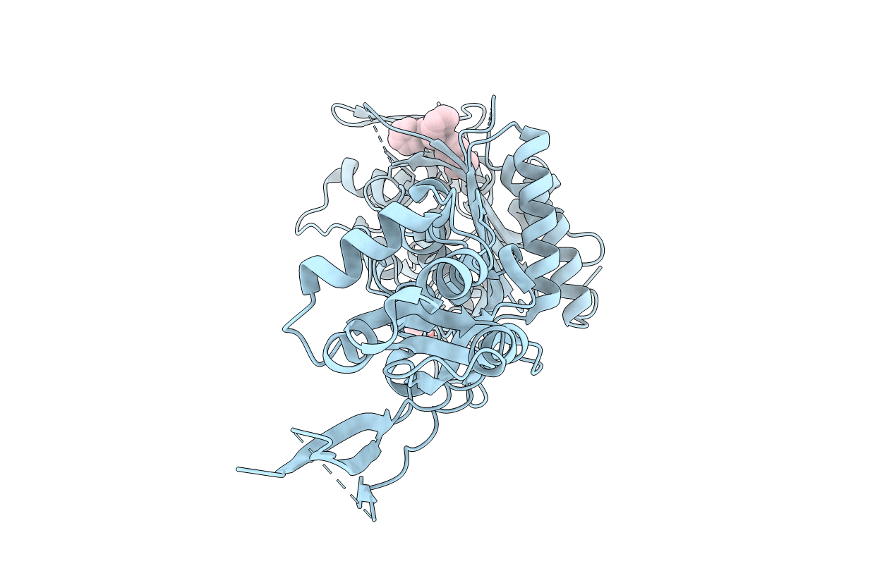
Organism: Pseudomonas aeruginosa
Method: X-RAY DIFFRACTION Resolution:1.41 Å Release Date: 2020-03-25 Classification: ANTIMICROBIAL PROTEIN Ligands: KDZ, KL8, ZN, CL, MG, GOL |
 |
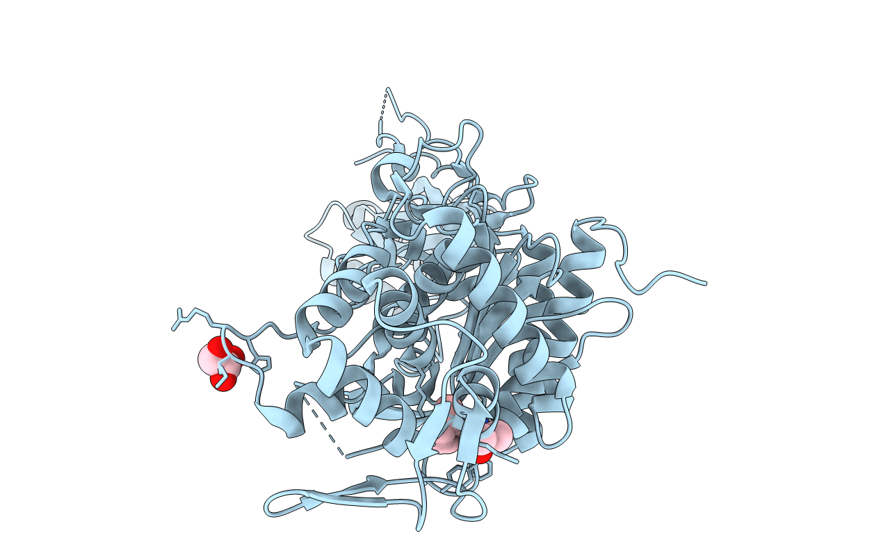
Organism: Klebsiella pneumoniae
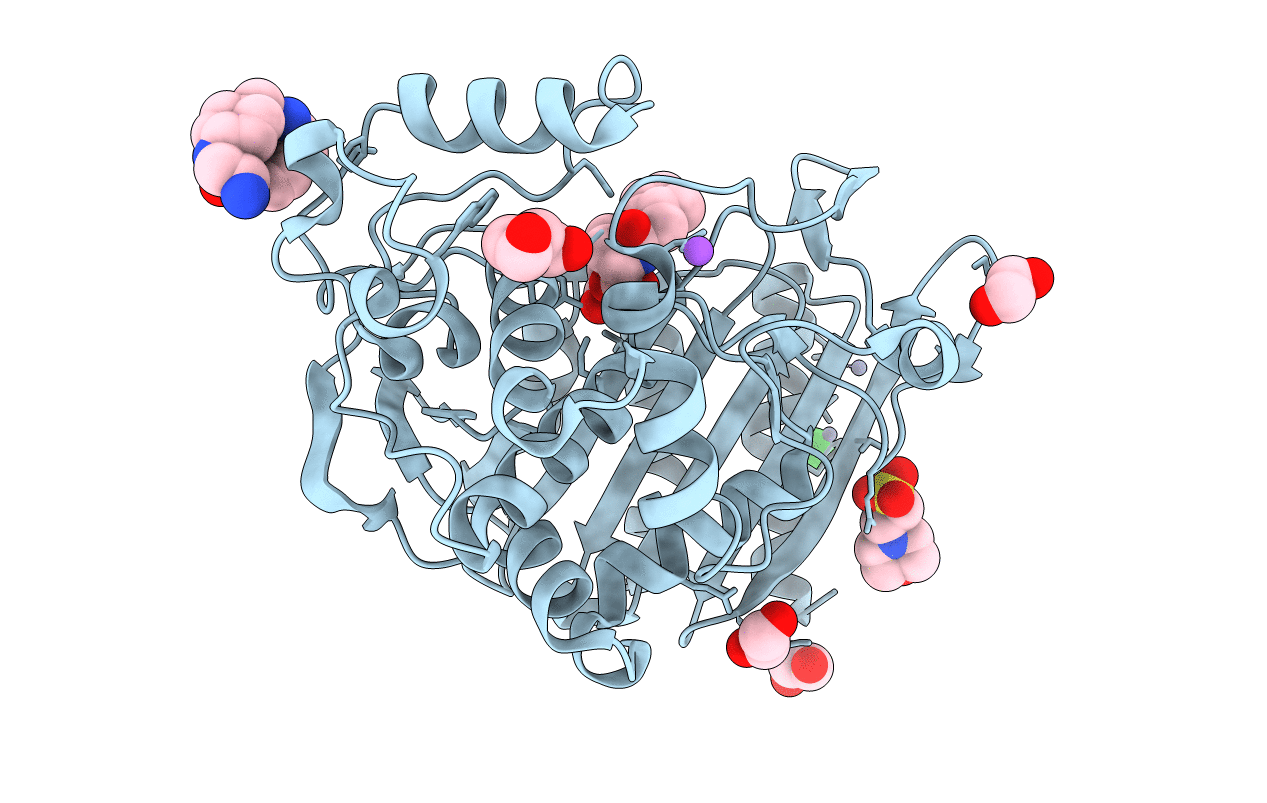
Method: X-RAY DIFFRACTION Resolution:0.99 Å Release Date: 2020-01-22 Classification: ANTIMICROBIAL PROTEIN Ligands: 4D6, GOL, SO4 |