Search Count: 18
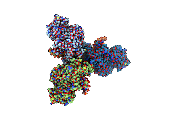 |
Organism: Homo sapiens, Synthetic construct
Method: X-RAY DIFFRACTION Resolution:2.26 Å Release Date: 2022-10-12 Classification: TRANSFERASE Ligands: MG, DG3 |
 |
Crystal Structure Of Pol Theta Polymerase Domain In Complex With Compound 5
Organism: Homo sapiens, Synthetic construct
Method: X-RAY DIFFRACTION Resolution:2.99 Å Release Date: 2022-10-12 Classification: TRANSFERASE Ligands: MG, DG3, K7X |
 |
Crystal Structure Of Pol Theta Polymerase Domain In Complex With Compound 22
Organism: Homo sapiens, Synthetic construct
Method: X-RAY DIFFRACTION Resolution:2.83 Å Release Date: 2022-10-12 Classification: TRANSFERASE Ligands: MG, DG3, K8I |
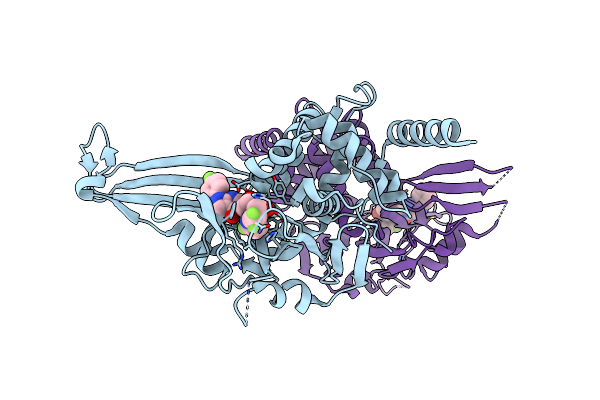 |
Crystal Structure Of Usp7 In Complex With The Non-Covalent Inhibitor, Ft671
Organism: Homo sapiens
Method: X-RAY DIFFRACTION Resolution:2.35 Å Release Date: 2017-10-18 Classification: HYDROLASE Ligands: 8WK |
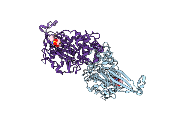 |
Organism: Homo sapiens
Method: X-RAY DIFFRACTION Resolution:2.33 Å Release Date: 2017-10-18 Classification: HYDROLASE Ligands: 8WN, EDO |
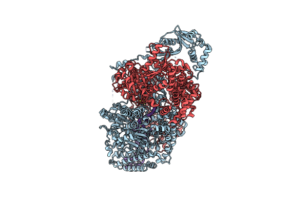 |
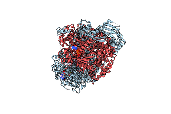
Organism: Bacillus subtilis subsp. subtilis str. 168, Synthetic construct
Method: X-RAY DIFFRACTION Resolution:3.24 Å Release Date: 2014-03-12 Classification: HYDROLASE/DNA Ligands: SF4 |
 |
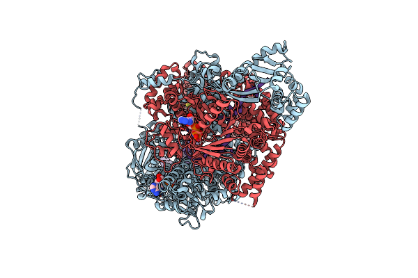
Organism: Bacillus subtilis subsp. subtilis str. 168, Synthetic construct
Method: X-RAY DIFFRACTION Resolution:2.80 Å Release Date: 2014-03-12 Classification: HYDROLASE/DNA Ligands: ANP, MG, SF4 |
 |
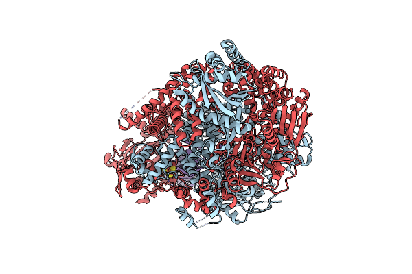
Organism: Bacillus subtilis subsp. subtilis str. 168, Synthetic construct
Method: X-RAY DIFFRACTION Resolution:3.00 Å Release Date: 2014-03-12 Classification: HYDROLASE/DNA Ligands: ANP, MG, SF4 |
 |
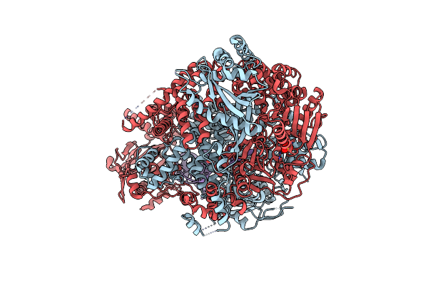
Organism: Bacillus subtilis
Method: X-RAY DIFFRACTION Resolution:3.20 Å Release Date: 2012-03-21 Classification: HYDROLASE/DNA Ligands: SF4 |
 |
Organism: Bacillus subtilis
Method: X-RAY DIFFRACTION Resolution:2.80 Å Release Date: 2012-03-21 Classification: HYDROLASE/DNA Ligands: SO4, EDO |
 |
Crystal Structure Of Mycobacterium Tuberculosis Glutamine Synthetase In Complex With A Purine Analogue Inhibitor.
Organism: Mycobacterium tuberculosis
Method: X-RAY DIFFRACTION Resolution:2.55 Å Release Date: 2009-09-01 Classification: LIGASE Ligands: 1AZ, CL |
 |
Crystal Structure Of Mycobacterium Tuberculosis Glutamine Synthetase In Complex With A Purine Analogue Inhibitor And L-Methionine-S- Sulfoximine Phosphate.
Organism: Mycobacterium tuberculosis
Method: X-RAY DIFFRACTION Resolution:2.20 Å Release Date: 2009-09-01 Classification: LIGASE Ligands: 1AZ, MG, P3S, PO4, CL, 1PE |
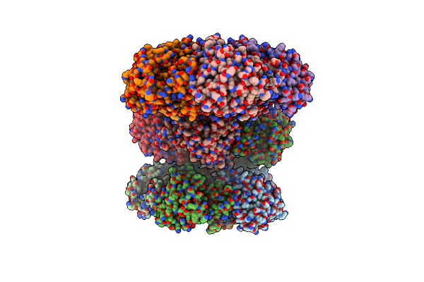 |
Organism: Canis familiaris
Method: X-RAY DIFFRACTION Resolution:3.00 Å Release Date: 2007-10-16 Classification: LIGASE Ligands: MG, CL |
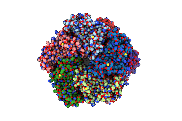 |
Crystal Structure Of Human Glutamine Synthetase In Complex With Adp And Methionine Sulfoximine Phosphate
Organism: Homo sapiens
Method: X-RAY DIFFRACTION Resolution:2.60 Å Release Date: 2007-07-03 Classification: LIGASE Ligands: MN, CL, ADP, P3S |
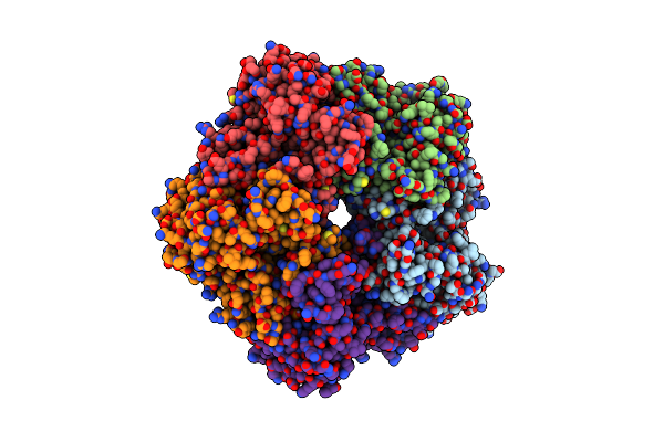 |
Crystal Structure Of Human Glutamine Synthetase In Complex With Adp And Phosphate
Organism: Homo sapiens
Method: X-RAY DIFFRACTION Resolution:2.05 Å Release Date: 2007-03-13 Classification: LIGASE Ligands: MN, PO4, CL, ADP, GOL |
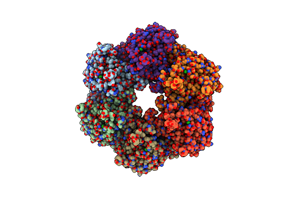 |
Crystal Structure Of Mycobacterium Tuberculosis Glutamine Synthetase In Complex With A Transition State Mimic
Organism: Mycobacterium tuberculosis
Method: X-RAY DIFFRACTION Resolution:2.10 Å Release Date: 2005-07-07 Classification: LIGASE Ligands: P3S, ADP, MG, CL |
 |
Organism: Escherichia coli
Method: X-RAY DIFFRACTION Release Date: 2004-03-02 Classification: STRUCTURAL GENOMICS, UNKNOWN FUNCTION |
 |
Structure Of The Double Mutant (L6M; F134M, Semet Form) Of Yqgf From Escherichia Coli, A Hypothetical Protein
Organism: Escherichia coli
Method: X-RAY DIFFRACTION Release Date: 2004-03-02 Classification: STRUCTURAL GENOMICS, UNKNOWN FUNCTION Ligands: SO4 |

