
Search Count: 18
 |
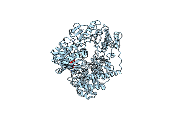
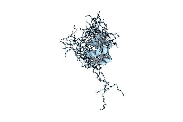
Semet-Derived Crystal Structure Of The N-Terminal Beta-Hairpin Docking (Bhd) Domain Of The Aerj Halogenase, From The Aeruginosin Biosynthetic Assembly Line
Organism: Microcystis aeruginosa nies-98
Method: X-RAY DIFFRACTION Resolution:1.68 Å Release Date: 2024-03-06 Classification: PROTEIN BINDING Ligands: CL |
 |
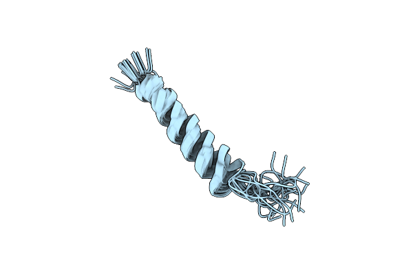
Native Crystal Structure Of The N-Terminal Beta-Hairpin Docking (Bhd) Domain Of The Aerj Halogenase, From The Aeruginosin Biosynthetic Assembly Line
Organism: Microcystis aeruginosa nies-98
Method: X-RAY DIFFRACTION Resolution:1.46 Å Release Date: 2024-03-06 Classification: PROTEIN BINDING Ligands: CL |
 |
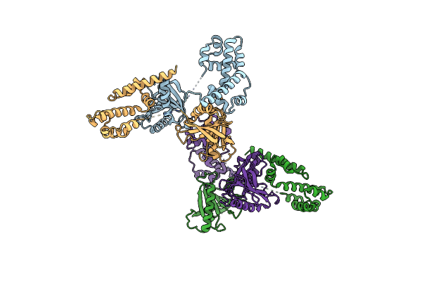
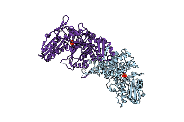
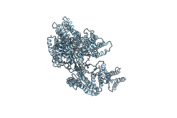
Discovery Of The Lanthipeptide Curvocidin And Structural Insights Into Its Trifunctional Synthetase Cuvl
Organism: Thermomonospora curvata
Method: X-RAY DIFFRACTION Resolution:2.68 Å Release Date: 2023-06-14 Classification: BIOSYNTHETIC PROTEIN Ligands: PO4, NO3 |
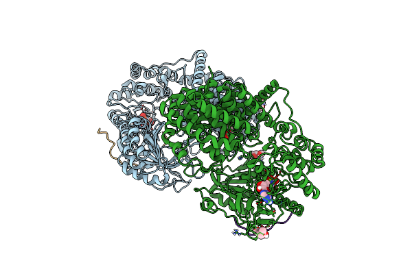 |
Discovery Of The Lanthipeptide Curvocidin And Structural Insights Into Its Trifunctional Synthetase Cuvl
Organism: Thermomonospora curvata
Method: X-RAY DIFFRACTION Resolution:2.87 Å Release Date: 2023-06-14 Classification: BIOSYNTHETIC PROTEIN Ligands: MPD, ANP, MG |
 |
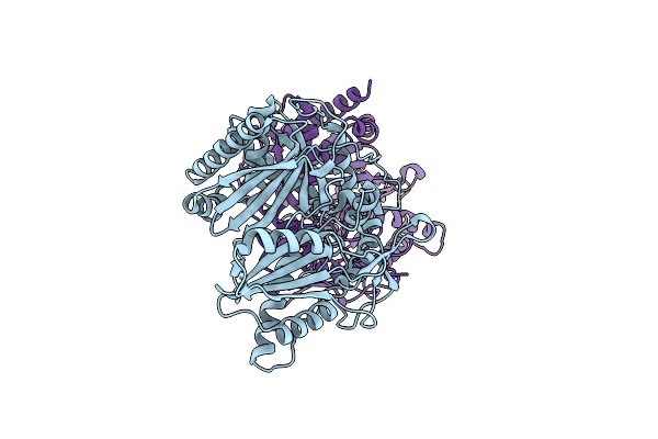
Crystal Structure Of The Albicidin Resistance Protein Stm3175 From Salmonella Typhimurium
Organism: Salmonella typhimurium
Method: X-RAY DIFFRACTION Resolution:3.60 Å Release Date: 2022-09-28 Classification: DNA BINDING PROTEIN |
 |
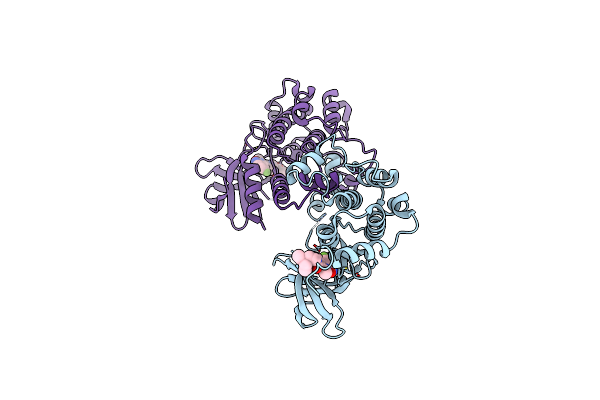
Crystal Structure Of The Epimerization Domain From The Third Module Of Tyrocidine Synthetase B, Tycb3(E)
Organism: Brevibacillus parabrevis
Method: X-RAY DIFFRACTION Resolution:2.40 Å Release Date: 2020-11-18 Classification: BIOSYNTHETIC PROTEIN |
 |
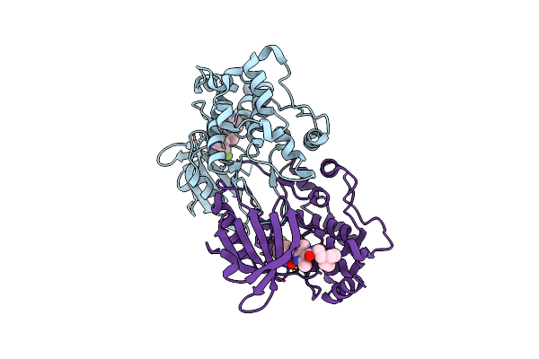
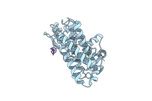
Crystal Structure Of S. Aureus Fabi In Complex With Nadph And Kalimantacin A (Batumin)
Organism: Staphylococcus aureus
Method: X-RAY DIFFRACTION Resolution:2.45 Å Release Date: 2020-04-01 Classification: BIOSYNTHETIC PROTEIN Ligands: NDP, KAL |
 |
Crystal Structure Of S. Aureus Fabi In Complex With Nadph And Kalimantacin B
Organism: Staphylococcus aureus
Method: X-RAY DIFFRACTION Resolution:2.55 Å Release Date: 2020-04-01 Classification: BIOSYNTHETIC PROTEIN Ligands: NDP, N5H |
 |
Crystal Structure Of Nonphosphorylated, Hpk1 Kinase Domain In Complex With Sunitinib In The Inactive State.
Organism: Homo sapiens
Method: X-RAY DIFFRACTION Resolution:2.17 Å Release Date: 2019-05-01 Classification: TRANSFERASE Ligands: B49 |
 |
Crystal Structure Of Diphosphorylated Hpk1 Kinase Domain In Complex With Sunitinib In The Active State.
Organism: Homo sapiens
Method: X-RAY DIFFRACTION Resolution:2.97 Å Release Date: 2019-05-01 Classification: SIGNALING PROTEIN Ligands: B49 |
 |
Crystal Structure Of Hpk1 Kinase Domain T165E,S171E Phosphomimetic Mutant In Complex With Sunitinib In The Inactive State.
Organism: Homo sapiens
Method: X-RAY DIFFRACTION Resolution:2.05 Å Release Date: 2019-05-01 Classification: TRANSFERASE Ligands: B49 |
 |
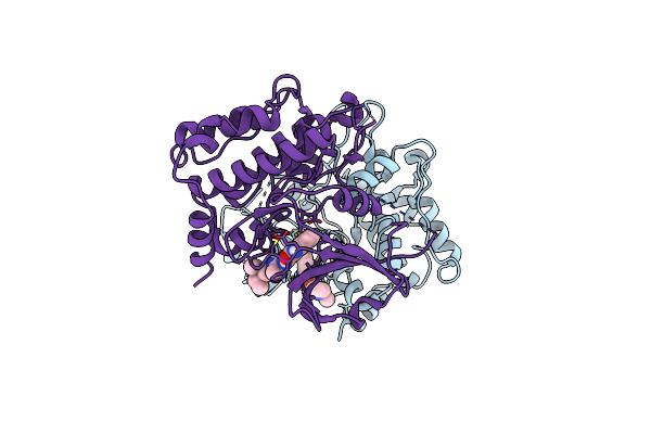
Molecular Basis For Condensation Domain-Mediated Chain Release From The Enacyloxin Polyketide Synthase
Organism: Burkholderia ambifaria (strain atcc baa-244 / ammd)
Method: X-RAY DIFFRACTION Resolution:2.00 Å Release Date: 2019-02-27 Classification: BIOSYNTHETIC PROTEIN Ligands: PO4 |
 |
Organism: Saccharomyces cerevisiae
Method: SOLUTION NMR Release Date: 2018-09-12 Classification: MEMBRANE PROTEIN |
 |
Organism: Burkholderia ambifaria ammd
Method: SOLUTION NMR Release Date: 2018-08-01 Classification: TRANSPORT PROTEIN |
 |
Organism: Phleum pratense
Method: SOLUTION NMR Release Date: 2014-03-26 Classification: PLANT PROTEIN |
 |
 |
Organism: Homo sapiens
Method: X-RAY DIFFRACTION Resolution:2.10 Å Release Date: 1999-05-18 Classification: COMPLEX (PROTO-ONCOGENE/PEPTIDE) Ligands: CA |
 |
Structure Of The N-Terminal Domain Of Cbl In Complex With Its Binding Site In Zap-70
Organism: Homo sapiens
Method: X-RAY DIFFRACTION Resolution:2.20 Å Release Date: 1999-04-27 Classification: SIGNAL TRANSDUCTION Ligands: CA |