
Search Count: 39
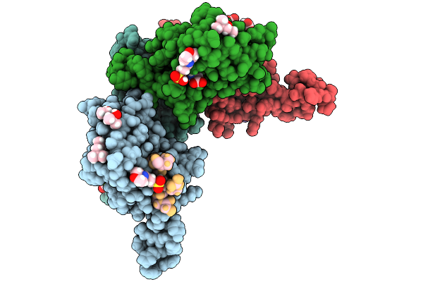 |
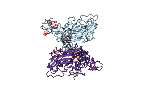
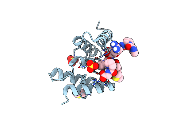
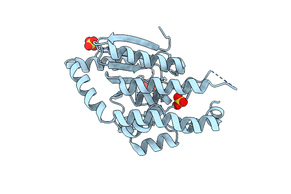
Peptide-Substrate-Binding (Psb) Domain Of Human Type I Collagen Prolyl 4-Hydroxylase Complexed With Pro-Pro-Gly-Pro-Arg-Gly-Pro-Pro-Gly.
Organism: Homo sapiens
Method: X-RAY DIFFRACTION Release Date: 2025-09-24 Classification: PROTEIN BINDING Ligands: MG, MPD, MPO, GLY |
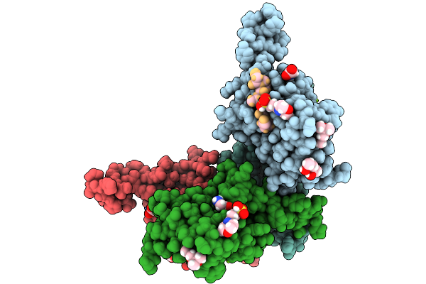 |
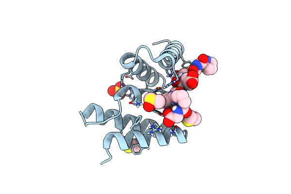
Peptide-Substrate-Binding (Psb) Domain Of Human Type I Collagen Prolyl 4-Hydroxylase Complexed With Pro-Hyp-Gly-Pro-Ala-Gly-Pro-Hyp-Gly.
Organism: Homo sapiens
Method: X-RAY DIFFRACTION Release Date: 2025-09-24 Classification: PROTEIN BINDING Ligands: GLY, MG, MPO, MPD |
 |
Peptide-Substrate-Binding (Psb) Domain Of Human Type I Collagen Prolyl 4-Hydroxylase Complexed With Pro-Pro-Gly-Pro-Ala-Gly-Pro-Pro-Gly.
Organism: Homo sapiens
Method: X-RAY DIFFRACTION Release Date: 2025-09-24 Classification: PROTEIN BINDING Ligands: GLY, MG, MPO, MPD |
 |
Peptide-Substrate-Binding (Psb) Domain Of Human Type Ii Collagen Prolyl 4-Hydroxylase Complexed With Pro-Hyp-Gly-Pro-Ala-Gly-Pro-Hyp-Gly.
Organism: Homo sapiens
Method: X-RAY DIFFRACTION Release Date: 2025-09-24 Classification: PROTEIN BINDING Ligands: GLY, SO4 |
 |
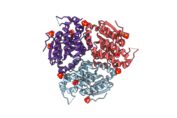
Crystal Structure Of Wild Type Perlecan Region 3 Construct I876-V1272 Construct Including One Laminin Iv-Like And Four Laminin Egf-Like Domains.
Organism: Mus musculus
Method: X-RAY DIFFRACTION Resolution:2.05 Å Release Date: 2025-04-02 Classification: STRUCTURAL PROTEIN Ligands: SIN, GOL |
 |
Crystal Structure Of Perlecan Region 3 Mutant (P1019L) Construct I876-V1272 Including One Laminin Iv-Like And Four Laminin Egf-Like Domains.
Organism: Mus musculus
Method: X-RAY DIFFRACTION Resolution:2.52 Å Release Date: 2025-04-02 Classification: STRUCTURAL PROTEIN Ligands: SIN |
 |
Organism: Mus musculus
Method: X-RAY DIFFRACTION Resolution:2.20 Å Release Date: 2024-07-10 Classification: STRUCTURAL PROTEIN |
 |
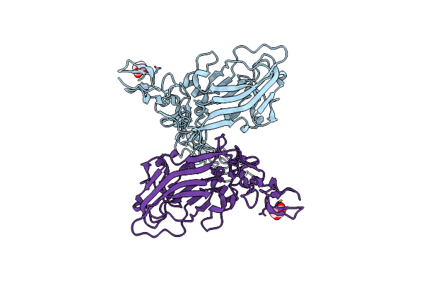
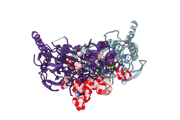
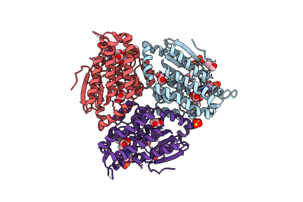
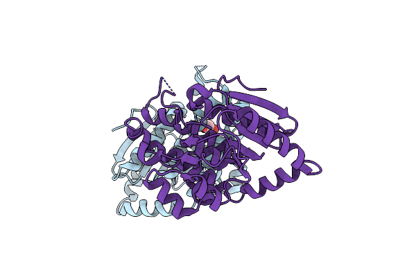
Crystal Structure Of The Heterodimeric Human C-P4H-Ii With Truncated Alpha Subunit (C-P4H-Ii Delta281)
Organism: Homo sapiens
Method: X-RAY DIFFRACTION Resolution:3.85 Å Release Date: 2022-11-09 Classification: HYDROLASE Ligands: SO4 |
 |
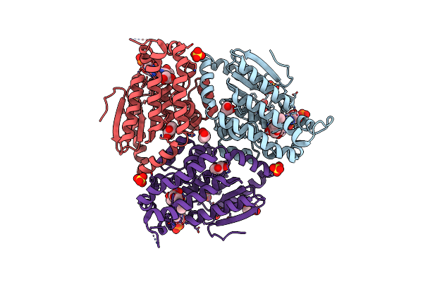
Organism: Homo sapiens
Method: X-RAY DIFFRACTION Resolution:2.25 Å Release Date: 2020-12-23 Classification: OXIDOREDUCTASE Ligands: OGA, FE2, TBU, GLY, CA, CL |
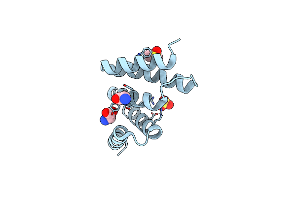 |
Crystal Structure Of An Unlignaded Peptide-Substrate-Binding Domain Of Human Type Ii Collagen Prolyl 4-Hydroxylase
Organism: Homo sapiens
Method: X-RAY DIFFRACTION Resolution:1.87 Å Release Date: 2018-09-12 Classification: HYDROLASE Ligands: SO4, DMS, GLY |
 |
Crystal Structure Of A Pro-9 Complexed Peptide-Substrate-Binding Domain Of Human Type Ii Collagen Prolyl 4-Hydroxylase
Organism: Homo sapiens, Synthetic construct
Method: X-RAY DIFFRACTION Resolution:2.00 Å Release Date: 2018-09-12 Classification: HYDROLASE Ligands: SO4, DMS |
 |
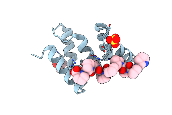
Crystal Structure Of Peptide-Substrate-Binding Domain Of Human Type Ii Collagen Prolyl 4-Hydroxylase Complex With Pro-Pro-Gly-Pro-Ala-Gly-Pro-Pro-Gly.
Organism: Homo sapiens, Synthetic construct
Method: X-RAY DIFFRACTION Resolution:1.48 Å Release Date: 2018-09-12 Classification: HYDROLASE Ligands: SO4, DMS |
 |
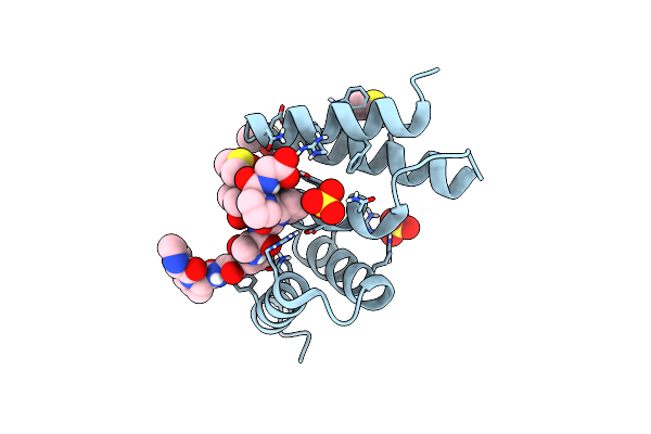
Crystal Structure The Peptide-Substrate-Binding Domain Of Human Type Ii Collagen Prolyl 4-Hydroxylase Complexed With Pro-Pro-Gly-Pro-Arg-Gly-Pro-Pro-Gly.
Organism: Homo sapiens, Synthetic construct
Method: X-RAY DIFFRACTION Resolution:1.55 Å Release Date: 2018-09-12 Classification: HYDROLASE Ligands: SO4, DMS |
 |
Crystal Structure The Peptide-Substrate-Binding Domain Of Human Type Ii Collagen Prolyl 4-Hydroxylase Complexed With Pro-Pro-Gly-Pro-Glu-Gly-Pro-Pro-Gly.
Organism: Homo sapiens, Synthetic construct
Method: X-RAY DIFFRACTION Resolution:1.68 Å Release Date: 2018-09-12 Classification: HYDROLASE Ligands: SO4, DMS |
 |
Organism: Saccharomyces cerevisiae (strain atcc 204508 / s288c)
Method: X-RAY DIFFRACTION Resolution:2.14 Å Release Date: 2015-11-11 Classification: ISOMERASE Ligands: SO4, CAA, GOL |
 |
Organism: Saccharomyces cerevisiae
Method: X-RAY DIFFRACTION Resolution:2.13 Å Release Date: 2015-11-11 Classification: ISOMERASE Ligands: SO4, CO8, GOL |
 |
Organism: Saccharomyces cerevisiae
Method: X-RAY DIFFRACTION Resolution:3.00 Å Release Date: 2015-11-11 Classification: ISOMERASE Ligands: SO4 |
 |
Organism: Saccharomyces cerevisiae
Method: X-RAY DIFFRACTION Resolution:2.10 Å Release Date: 2015-11-11 Classification: ISOMERASE Ligands: GOL, SO4 |
 |
Crystal Structure Of Yeast Enoyl-Coa Isomerase Helix-10 Deletion (Sceci2-H10) Mutant
Organism: Saccharomyces cerevisiae
Method: X-RAY DIFFRACTION Resolution:1.81 Å Release Date: 2015-11-11 Classification: ISOMERASE Ligands: GOL |
 |
Cdsd - The Structural Protein Of The Type Iii Secretion System Of Chlamydia Trachomatis: C-Terminal Domain
Organism: Chlamydia trachomatis
Method: X-RAY DIFFRACTION Resolution:2.00 Å Release Date: 2015-07-01 Classification: STRUCTURAL PROTEIN Ligands: SO4 |