
Search Count: 21
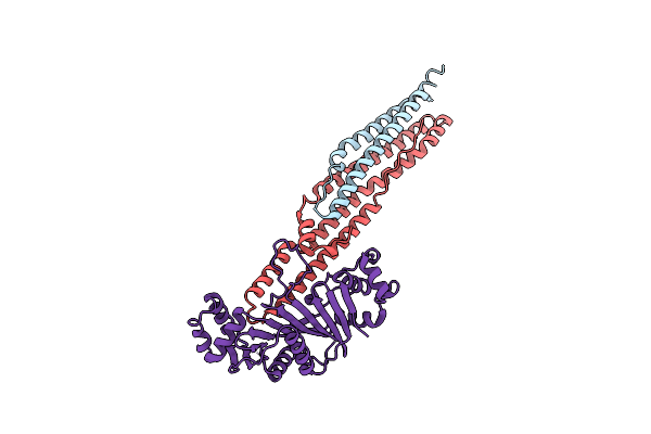 |
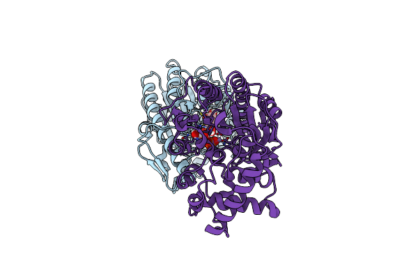
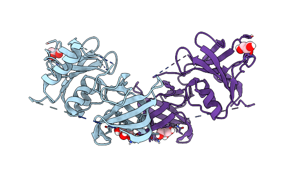
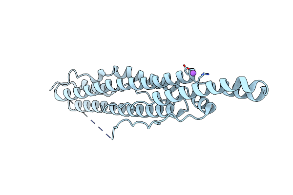
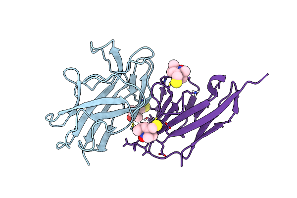
Structure Of Pe25-Ppe41(A124L) In Complex With Espg5 Chaperone From The Type Vii (Esx-5) Secretion System
Organism: Mycobacterium tuberculosis (strain atcc 25618 / h37rv), Mycobacterium marinum (strain atcc baa-535 / m)
Method: X-RAY DIFFRACTION Resolution:2.40 Å Release Date: 2021-01-27 Classification: PROTEIN TRANSPORT Ligands: CL |
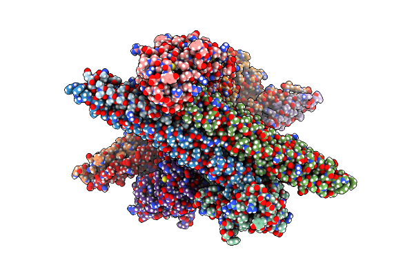 |
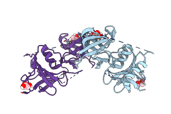
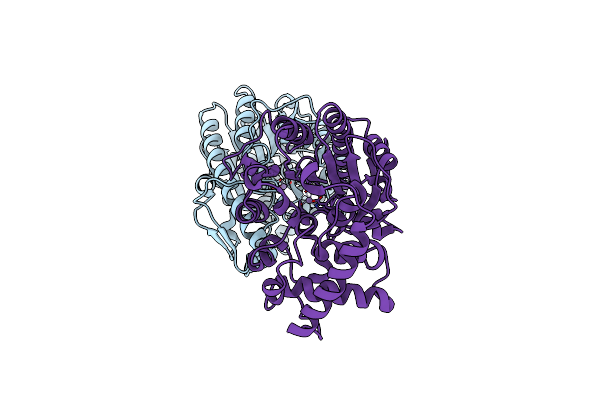
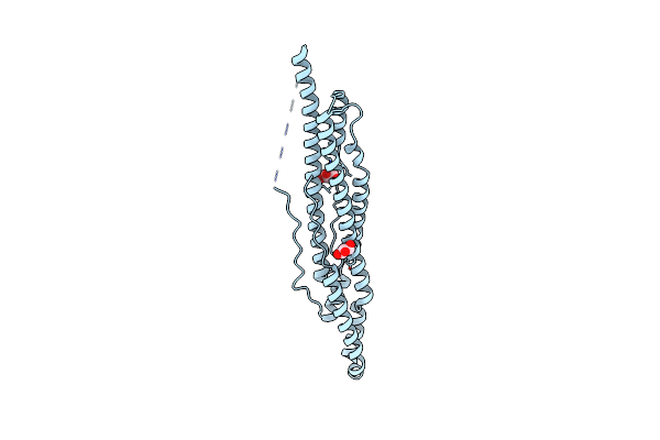
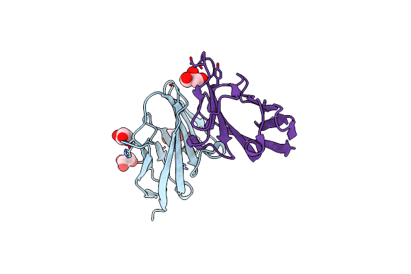
Structure Of Pe5-Ppe4-Espg3 Complex From The Type Vii (Esx-3) Secretion System, Space Group P212121
Organism: Mycobacterium tuberculosis (strain atcc 25618 / h37rv), Mycobacterium marinum (strain atcc baa-535 / m)
Method: X-RAY DIFFRACTION Resolution:3.00 Å Release Date: 2020-01-29 Classification: PROTEIN TRANSPORT |
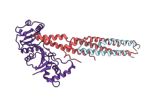 |
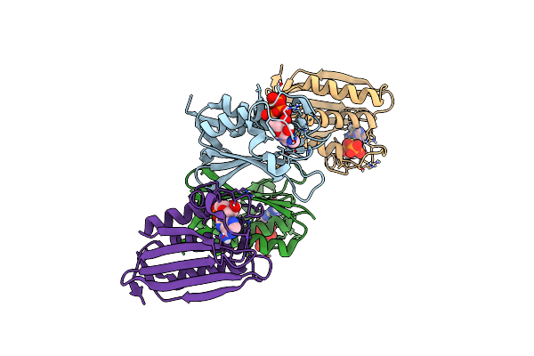
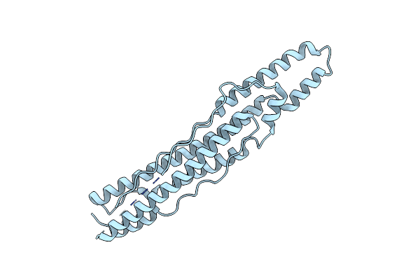
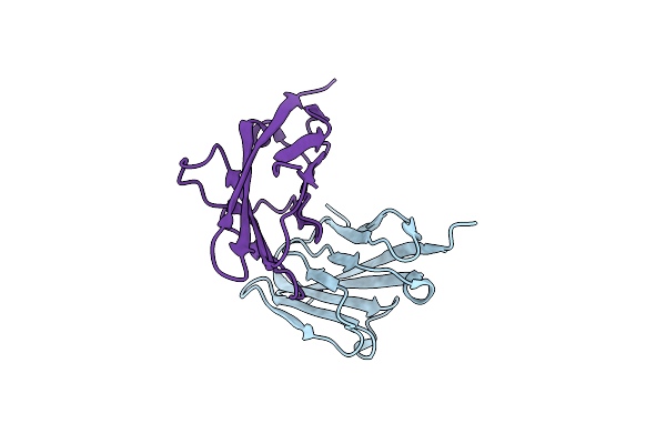
Structure Of Pe5-Ppe4-Espg3 Complex From The Type Vii (Esx-3) Secretion System, Space Group I422
Organism: Mycobacterium tuberculosis (strain atcc 25618 / h37rv), Mycobacterium marinum (strain atcc baa-535 / m)
Method: X-RAY DIFFRACTION Resolution:3.30 Å Release Date: 2020-01-29 Classification: PROTEIN TRANSPORT |
 |
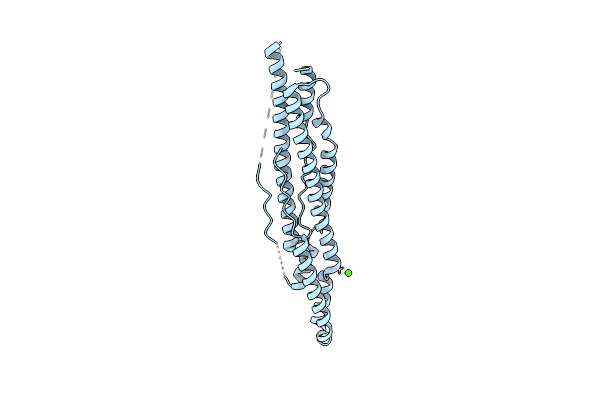
Organism: Enterococcus faecium tx0133a04
Method: X-RAY DIFFRACTION Resolution:1.50 Å Release Date: 2019-07-03 Classification: TRANSFERASE Ligands: GOL |
 |
Organism: Streptococcus pyogenes
Method: X-RAY DIFFRACTION Resolution:1.78 Å Release Date: 2019-05-01 Classification: HYDROLASE Ligands: ACT, ZN, EDO, IMD |
 |
Organism: Enterococcus faecalis (strain atcc 700802 / v583)
Method: X-RAY DIFFRACTION Resolution:2.18 Å Release Date: 2019-03-27 Classification: TRANSFERASE Ligands: GOL, PO4, CL, NA |
 |
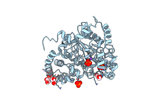
Streptococcus Pyogenes Phosphoglycerol Transferase Gach In Complex With Sn-Glycerol-1-Phosphate, Crystal Form 1
Organism: Streptococcus pyogenes serotype m1
Method: X-RAY DIFFRACTION Resolution:2.00 Å Release Date: 2018-07-11 Classification: TRANSFERASE Ligands: MN, 1GP, CA |
 |
Streptococcus Pyogenes Phosphoglycerol Transferase Gach In Complex With Sn-Glycerol-1-Phosphate
Organism: Streptococcus pyogenes mgas5005
Method: X-RAY DIFFRACTION Resolution:1.49 Å Release Date: 2018-06-06 Classification: TRANSFERASE Ligands: MN, 1GP, CA |
 |
Phage-Associated Cell Wall Hydrolase Plypy From Streptococcus Pyogenes, Space Group P6522
Organism: Streptococcus pyogenes mgas5005
Method: X-RAY DIFFRACTION Resolution:2.64 Å Release Date: 2017-12-27 Classification: HYDROLASE Ligands: GOL |
 |
Phage-Associated Cell Wall Hydrolase Plypy From Streptococcus Pyogenes, Space Group P3121
Organism: Streptococcus pyogenes mgas5005
Method: X-RAY DIFFRACTION Resolution:1.97 Å Release Date: 2017-12-27 Classification: HYDROLASE Ligands: GOL |
 |
Organism: Streptococcus pyogenes mgas5005
Method: X-RAY DIFFRACTION Resolution:2.00 Å Release Date: 2017-12-20 Classification: TRANSFERASE Ligands: MN |
 |
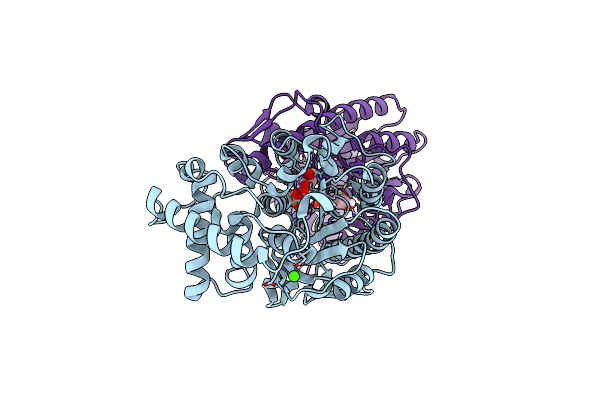
Organism: Mycobacterium hassiacum (strain dsm 44199 / cip 105218 / jcm 12690 / 3849)
Method: X-RAY DIFFRACTION Resolution:1.55 Å Release Date: 2017-11-22 Classification: TRANSFERASE Ligands: ATP, MG |
 |
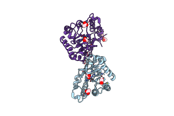
Structure Of Pe-Ppe Domains Of Esx-1 Secreted Protein Espb, C2221 In Presence Of Ca
Organism: Mycobacterium tuberculosis
Method: X-RAY DIFFRACTION Resolution:1.82 Å Release Date: 2015-02-18 Classification: PROTEIN TRANSPORT Ligands: CA |
 |
Organism: Mycobacterium tuberculosis
Method: X-RAY DIFFRACTION Resolution:2.14 Å Release Date: 2015-02-18 Classification: PROTEIN TRANSPORT Ligands: CL, NA |
 |
Organism: Mycobacterium tuberculosis
Method: X-RAY DIFFRACTION Resolution:1.50 Å Release Date: 2015-02-18 Classification: PROTEIN TRANSPORT Ligands: CL, GOL |
 |
Organism: Mycobacterium tuberculosis
Method: X-RAY DIFFRACTION Resolution:3.04 Å Release Date: 2015-02-18 Classification: PROTEIN TRANSPORT |
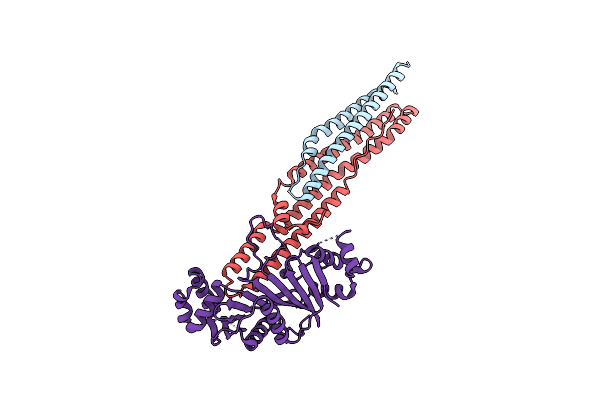 |
Structure Of The Mycobacterium Tuberculosis Type Vii Secretion System Chaperone Espg5 In Complex With Pe25-Ppe41 Dimer
Organism: Mycobacterium tuberculosis
Method: X-RAY DIFFRACTION Resolution:2.60 Å Release Date: 2014-05-28 Classification: PROTEIN TRANSPORT |
 |
Structural Model For The Complex Between The Dr Adhesins And Carcinoembryonic Antigen (Cea)
Organism: Escherichia coli, Homo sapiens
Method: SOLUTION NMR Release Date: 2008-01-08 Classification: CELL ADHESION Ligands: MTN |
 |
Crystal Structure Of The N-Terminal Domain Of Carcinoembryonic Antigen (Cea)
Organism: Homo sapiens
Method: X-RAY DIFFRACTION Resolution:1.95 Å Release Date: 2008-01-01 Classification: CELL ADHESION Ligands: CL, GOL |
 |
Crystal Structure Of The V39C Mutant Of The N-Terminal Domain Of Carcinoembryonic Antigen (Cea)
Organism: Homo sapiens
Method: X-RAY DIFFRACTION Resolution:2.90 Å Release Date: 2008-01-01 Classification: CELL ADHESION |