
Search Count: 16
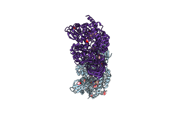 |
Erap1 In Complex With 1-[2-(3-Oxo-3,4-Dihydro-2H-1,4-Benzothiazin-4-Yl)Acetamido]Cyclohexane-1-Carboxylic Acid
Organism: Homo sapiens
Method: X-RAY DIFFRACTION Resolution:1.72 Å Release Date: 2025-01-22 Classification: PEPTIDE BINDING PROTEIN Ligands: ZN, B3P, EDO, BR, A1IMK |
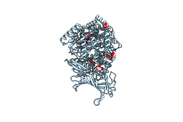 |
Erap1 In Complex With 1-[2-(6-Bromo-3-Oxo-3,4-Dihydro-2H-1,4-Benzoxazin-4-Yl)Acetamido]-4,4-Difluorocyclohexane-1-Carboxylic Acid
Organism: Homo sapiens
Method: X-RAY DIFFRACTION Resolution:1.35 Å Release Date: 2025-01-22 Classification: PEPTIDE BINDING PROTEIN Ligands: ZN, PO4, EDO, A1IMJ |
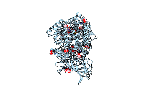 |
Erap1 In Complex With 1-[2-(6-Chloro-3-Oxo-3,4-Dihydro-2H-1,4-Benzothiazin-4-Yl)Acetamido]Cyclohexane-1-Carboxylic Acid
Organism: Homo sapiens
Method: X-RAY DIFFRACTION Resolution:1.33 Å Release Date: 2025-01-22 Classification: PEPTIDE BINDING PROTEIN Ligands: ZN, MLT, EDO, A1IMM |
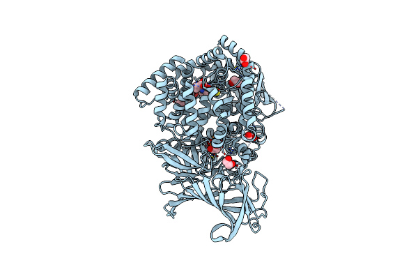 |
Erap1 In Complex With 1-[2-(2-Oxo-5-Phenyl-2,3-Dihydro-1,3-Benzothiazol-3-Yl)Acetamido]Cyclohexane-1-Carboxylic Acid
Organism: Homo sapiens
Method: X-RAY DIFFRACTION Resolution:1.37 Å Release Date: 2025-01-22 Classification: PEPTIDE BINDING PROTEIN Ligands: ZN, PO4, EDO, A1IML |
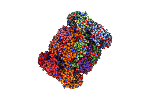 |
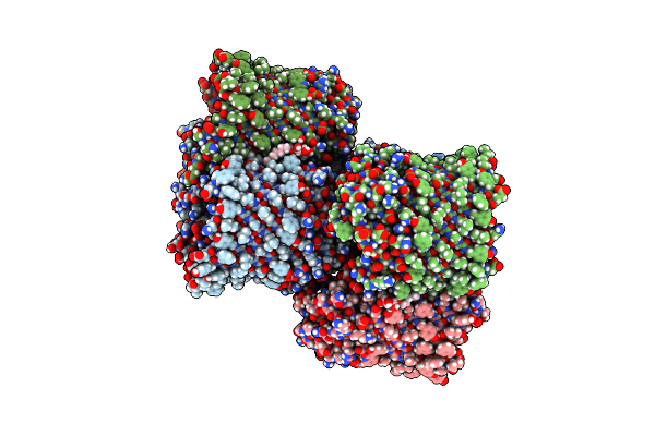
Leishmania Tarentolae Proteasome 20S Subunit In Complex With 1-Benzyl-N-(3-(Cyclopropylcarbamoyl)Phenyl)-6-Oxo-1,6-Dihydropyridazine-3-Carboxamide
Organism: Leishmania tarentolae
Method: ELECTRON MICROSCOPY Resolution:2.59 Å Release Date: 2023-08-09 Classification: UNKNOWN FUNCTION Ligands: VYW |
 |
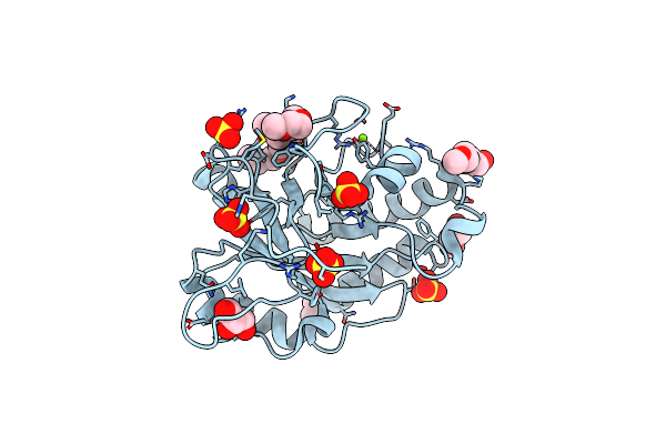
Human Endoplasmic Reticulum Aminopeptidase 1 (Erap1) In Complex With (4Ar,5S,6R,8S,8Ar)-5-(2-(Furan-3-Yl)Ethyl)-8-Hydroxy-5,6,8A-Trimethyl-3,4,4A,5,6,7,8,8A-Octahydronaphthalene-1-Carboxylic Acid
Organism: Homo sapiens
Method: X-RAY DIFFRACTION Resolution:1.67 Å Release Date: 2020-03-18 Classification: HYDROLASE Ligands: ZN, MLT, EDO, MNZ |
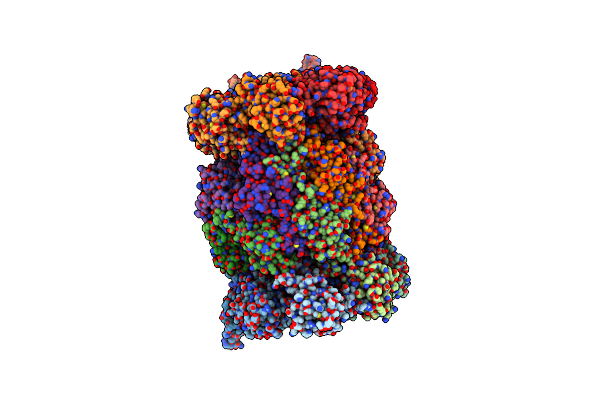 |
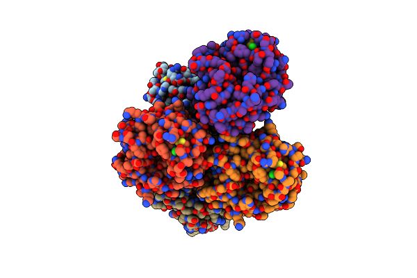
Organism: Leishmania tarentolae
Method: ELECTRON MICROSCOPY Resolution:2.80 Å Release Date: 2019-04-17 Classification: HYDROLASE Ligands: J6E |
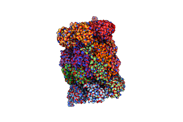 |
Organism: Leishmania tarentolae
Method: ELECTRON MICROSCOPY Resolution:3.30 Å Release Date: 2019-04-17 Classification: HYDROLASE |
 |
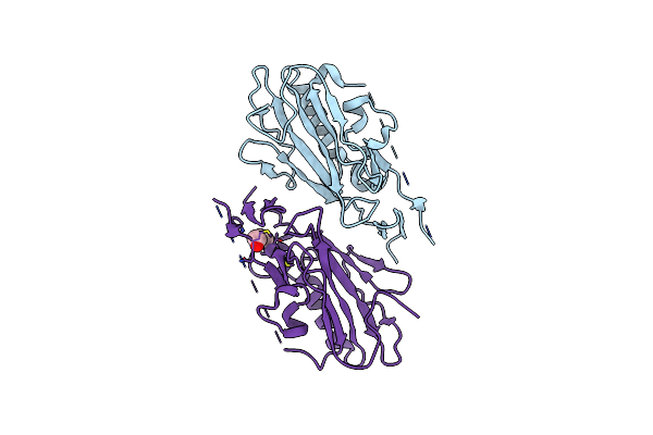
Organism: Homo sapiens
Method: X-RAY DIFFRACTION Resolution:2.70 Å Release Date: 2016-02-17 Classification: IMMUNE SYSTEM |
 |
Organism: Homo sapiens
Method: X-RAY DIFFRACTION Resolution:3.45 Å Release Date: 2016-02-17 Classification: HORMONE |
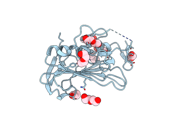 |
Organism: Streptococcus pyogenes serotype m1
Method: X-RAY DIFFRACTION Resolution:1.70 Å Release Date: 2011-07-20 Classification: HYDROLASE Ligands: PEG |
 |
Organism: Streptococcus pyogenes serotype m1
Method: X-RAY DIFFRACTION Resolution:2.49 Å Release Date: 2011-07-20 Classification: HYDROLASE Ligands: PEG |
 |
Organism: Escherichia coli
Method: X-RAY DIFFRACTION Resolution:3.01 Å Release Date: 2010-12-08 Classification: MEMBRANE PROTEIN Ligands: BOG |
 |
Organism: Aspergillus fumigatus
Method: X-RAY DIFFRACTION Resolution:1.70 Å Release Date: 2010-09-08 Classification: HYDROLASE Ligands: CL |
 |
Organism: Bacillus anthracis
Method: X-RAY DIFFRACTION Resolution:1.40 Å Release Date: 2009-06-23 Classification: HYDROLASE Ligands: 15P, SO4, GOL, MG |
 |
Crystal Structure Of Human Rhoa In Complex With Dh/Ph Fragment Of Pdzrhogef
Organism: Homo sapiens
Method: X-RAY DIFFRACTION Resolution:2.50 Å Release Date: 2004-12-14 Classification: SIGNALING PROTEIN ACTIVATOR/SIGNALING PROTEIN |