
Search Count: 31
 |
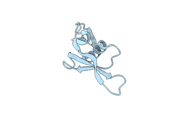
Organism: Pseudomonas aeruginosa
Method: SOLUTION NMR Release Date: 2025-06-18 Classification: VIRAL PROTEIN |
 |
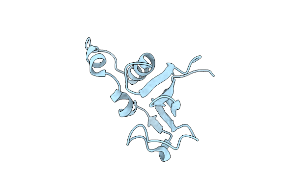
Organism: Pseudomonas aeruginosa
Method: X-RAY DIFFRACTION Release Date: 2025-06-18 Classification: VIRAL PROTEIN |
 |
Organism: Human immunodeficiency virus 1, Macaca
Method: ELECTRON MICROSCOPY Release Date: 2024-09-11 Classification: VIRAL PROTEIN Ligands: NAG |
 |
Organism: Macaca, Human immunodeficiency virus 1
Method: ELECTRON MICROSCOPY Release Date: 2024-05-29 Classification: VIRAL PROTEIN Ligands: NAG |
 |
Organism: Pseudomonas phage dms3
Method: X-RAY DIFFRACTION Resolution:1.33 Å Release Date: 2024-05-08 Classification: VIRAL PROTEIN |
 |
Organism: Bacillus cereus (strain vd045)
Method: X-RAY DIFFRACTION Resolution:2.00 Å Release Date: 2024-01-31 Classification: UNKNOWN FUNCTION |
 |
Organism: Macaca mulatta
Method: X-RAY DIFFRACTION Release Date: 2023-12-13 Classification: VIRAL PROTEIN/IMMUNE SYSTEM |
 |
Organism: Pseudomonas phage jbd88a
Method: X-RAY DIFFRACTION Resolution:1.23 Å Release Date: 2023-09-20 Classification: VIRAL PROTEIN |
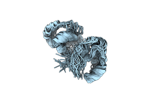 |
Organism: Pseudomonas citronellolis
Method: SOLUTION NMR Release Date: 2022-02-09 Classification: VIRAL PROTEIN |
 |
Organism: Haemophilus influenzae (strain atcc 51907 / dsm 11121 / kw20 / rd)
Method: X-RAY DIFFRACTION Resolution:2.70 Å Release Date: 2021-09-29 Classification: UNKNOWN FUNCTION Ligands: CL |
 |
Organism: Pseudomonas aeruginosa
Method: SOLUTION NMR Release Date: 2020-08-26 Classification: HYDROLASE INHIBITOR |
 |
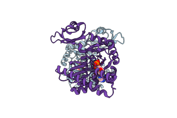
Organism: Homo sapiens
Method: X-RAY DIFFRACTION Resolution:2.90 Å Release Date: 2020-03-04 Classification: OXIDOREDUCTASE |
 |
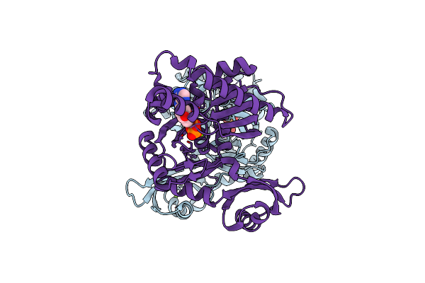
Organism: Homo sapiens
Method: X-RAY DIFFRACTION Resolution:3.00 Å Release Date: 2020-03-04 Classification: OXIDOREDUCTASE Ligands: NAD |
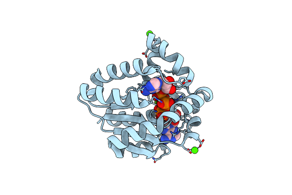 |
Organism: Bacillus subtilis
Method: X-RAY DIFFRACTION Resolution:2.00 Å Release Date: 2018-02-28 Classification: TRANSFERASE Ligands: ZN, MG, AP5, CA |
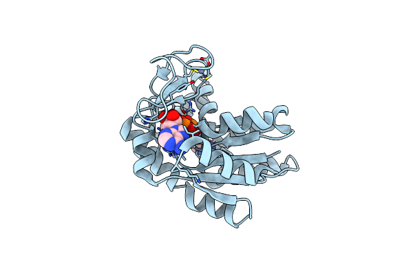 |
Organism: Sporosarcina globispora
Method: X-RAY DIFFRACTION Resolution:2.10 Å Release Date: 2018-02-28 Classification: TRANSFERASE Ligands: ZN, AP5 |
 |
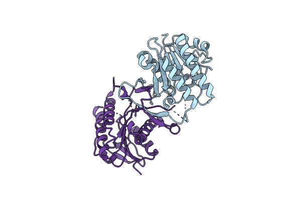
Organism: Pseudomonas aeruginosa (strain atcc 15692 / pao1 / 1c / prs 101 / lmg 12228), Pseudomonas aeruginosa
Method: X-RAY DIFFRACTION Resolution:2.40 Å Release Date: 2016-04-27 Classification: PEPTIDE BINDING PROTEIN Ligands: SO4, CL, NA, MG, ATP |
 |
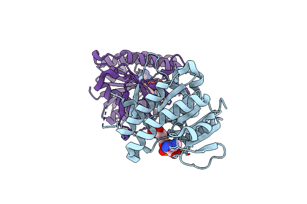
Organism: Pseudomonas aeruginosa (strain atcc 15692 / pao1 / 1c / prs 101 / lmg 12228)
Method: X-RAY DIFFRACTION Resolution:2.40 Å Release Date: 2016-04-27 Classification: PEPTIDE BINDING PROTEIN Ligands: ADP, MG |
 |
Organism: Pseudomonas aeruginosa
Method: X-RAY DIFFRACTION Resolution:2.50 Å Release Date: 2016-04-27 Classification: PEPTIDE BINDING PROTEIN Ligands: ADP, MG |
 |
Organism: Pseudomonas aeruginosa (strain atcc 15692 / pao1 / 1c / prs 101 / lmg 12228)
Method: X-RAY DIFFRACTION Resolution:3.50 Å Release Date: 2016-04-27 Classification: PEPTIDE BINDING PROTEIN Ligands: ANP, MG |
 |
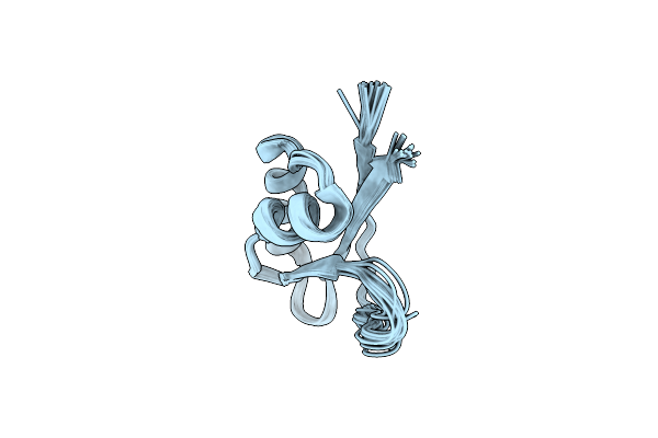
Organism: Pseudomonas aeruginosa
Method: SOLUTION NMR Release Date: 2011-12-21 Classification: STRUCTURAL PROTEIN |