
Search Count: 13
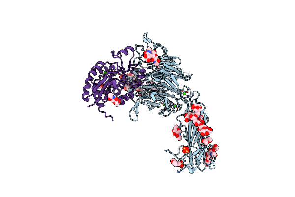 |
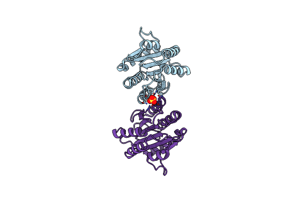
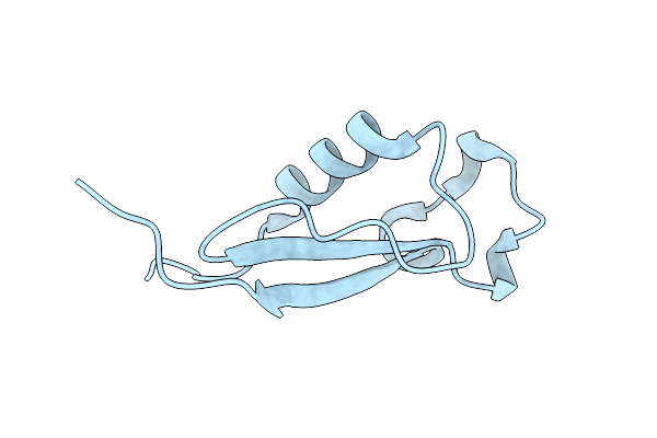
Organism: Homo sapiens
Method: X-RAY DIFFRACTION Resolution:2.25 Å Release Date: 2017-01-25 Classification: CELL ADHESION Ligands: CA, NAG, SO4, MES, PEG |
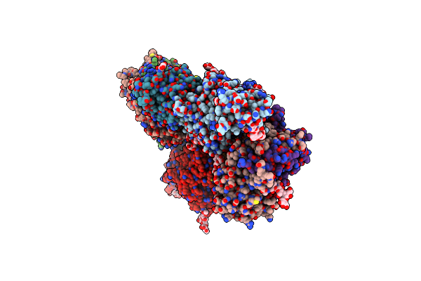 |
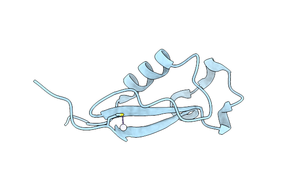
Organism: Homo sapiens
Method: X-RAY DIFFRACTION Resolution:3.49 Å Release Date: 2017-01-25 Classification: CELL ADHESION Ligands: CA, NAG, MN |
 |
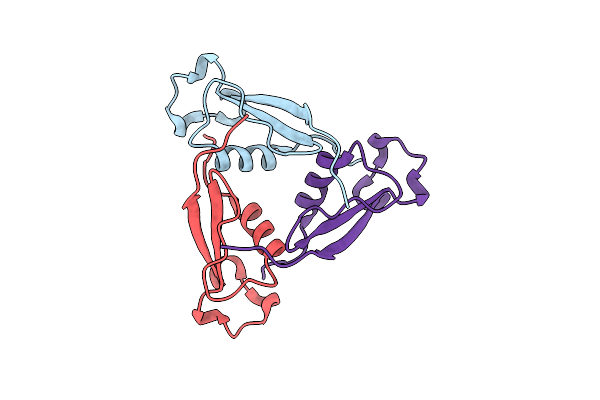
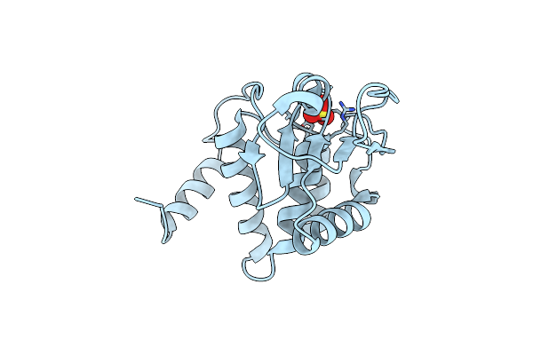
Organism: Homo sapiens
Method: X-RAY DIFFRACTION Resolution:2.30 Å Release Date: 2014-12-31 Classification: HYDROLASE Ligands: SO4, BME |
 |
Organism: Plasmodium falciparum
Method: X-RAY DIFFRACTION Resolution:2.20 Å Release Date: 2012-12-26 Classification: CELL ADHESION Ligands: CL |
 |
Organism: Plasmodium falciparum
Method: X-RAY DIFFRACTION Resolution:2.25 Å Release Date: 2012-12-26 Classification: CELL ADHESION Ligands: SO4 |
 |
Organism: Plasmodium vivax
Method: X-RAY DIFFRACTION Resolution:2.24 Å Release Date: 2012-12-26 Classification: CELL ADHESION Ligands: MG, CL |
 |
Organism: Plasmodium vivax
Method: X-RAY DIFFRACTION Resolution:2.20 Å Release Date: 2012-12-26 Classification: CELL ADHESION Ligands: MN, CL |
 |
Organism: Plasmodium vivax
Method: X-RAY DIFFRACTION Resolution:2.19 Å Release Date: 2012-12-26 Classification: CELL ADHESION Ligands: MG, CL, NA |
 |
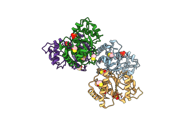
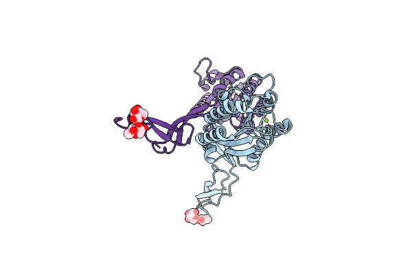
Organism: Plasmodium falciparum
Method: X-RAY DIFFRACTION Resolution:1.70 Å Release Date: 2012-05-09 Classification: CELL INVASION |
 |
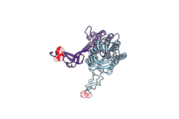
Crystal Structure Of Circumsporozoite Protein Atsr Domain, R32 Platinum-Bound Form
Organism: Plasmodium falciparum
Method: X-RAY DIFFRACTION Resolution:1.85 Å Release Date: 2012-05-09 Classification: CELL INVASION Ligands: PT |
 |
Organism: Plasmodium falciparum
Method: X-RAY DIFFRACTION Resolution:2.04 Å Release Date: 2012-05-09 Classification: CELL INVASION |
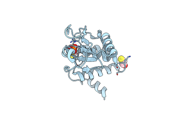 |
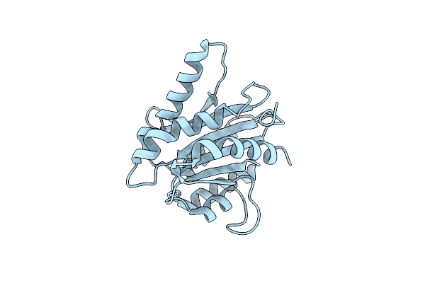
High Resolution Crystal Structure Of The Vaccinia Virus Dual-Specificity Phosphatase Vh1
Organism: Vaccinia virus
Method: X-RAY DIFFRACTION Resolution:1.32 Å Release Date: 2009-02-10 Classification: HYDROLASE Ligands: PO4, BME |
 |
Organism: Vaccinia virus
Method: X-RAY DIFFRACTION Resolution:1.95 Å Release Date: 2008-09-30 Classification: HYDROLASE Ligands: SO4 |