Planned Maintenance: Some services may turn out to be unavailable from 15th January, 2026 to 16th January, 2026. We apologize for the inconvenience!

Planned Maintenance: Some services may turn out to be unavailable from 15th January, 2026 to 16th January, 2026. We apologize for the inconvenience!
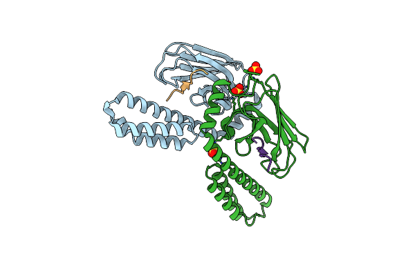 |
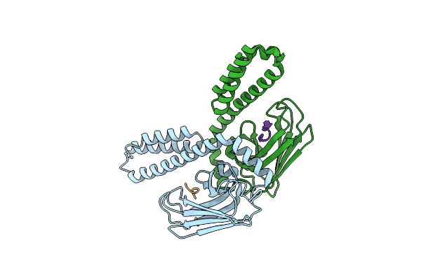
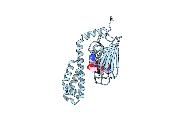
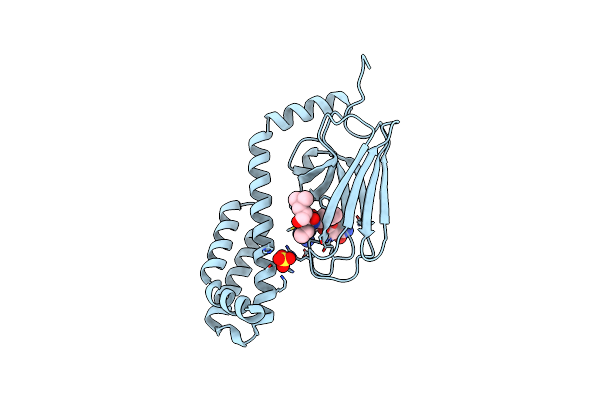
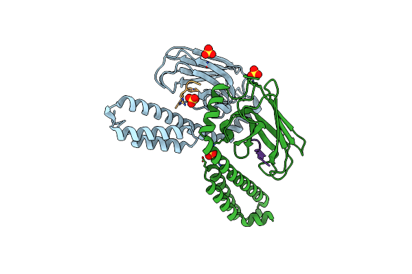
Crystal Structure Of The Substrate Binding Domain Of E.Coli Dnak In Complex With Bovine Bac7(1-16)
Organism: Escherichia coli, Bos taurus
Method: X-RAY DIFFRACTION Resolution:1.80 Å Release Date: 2013-11-13 Classification: CHAPERONE/Antibiotic Ligands: SO4 |
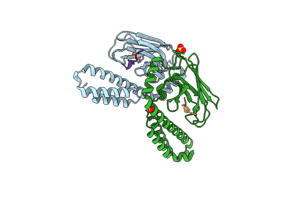 |
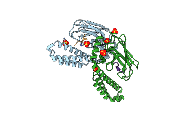
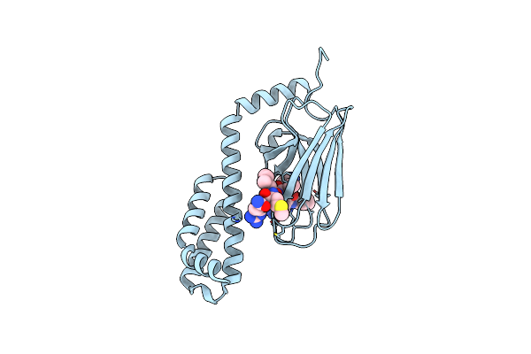
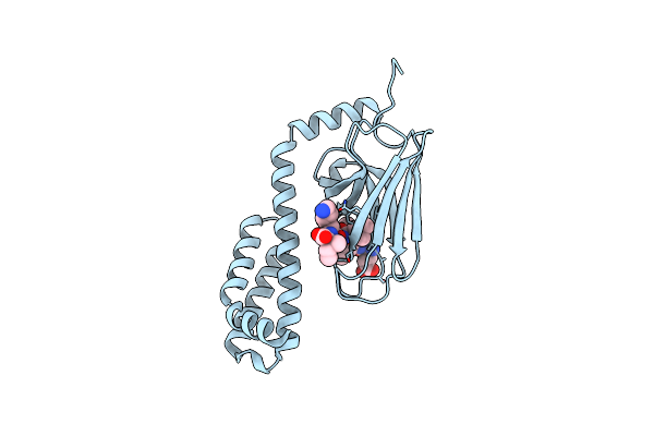
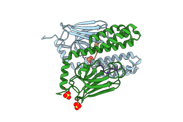
Crystal Structure Of The Substrate Binding Domain Of E.Coli Dnak In Complex With Bovine Bac7(15-28)
Organism: Escherichia coli, Bos taurus
Method: X-RAY DIFFRACTION Resolution:1.95 Å Release Date: 2013-11-13 Classification: CHAPERONE/Antibiotic Ligands: SO4 |
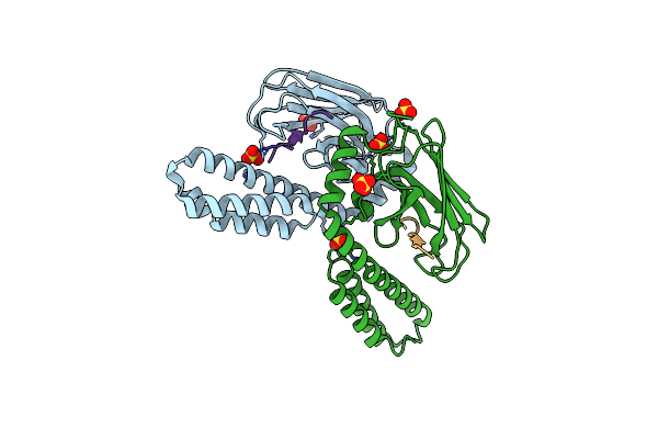 |
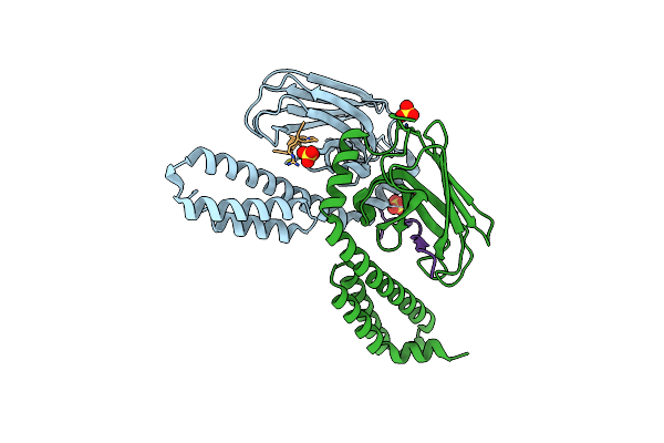
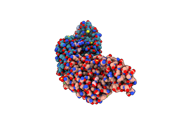
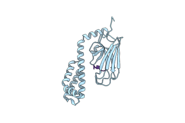
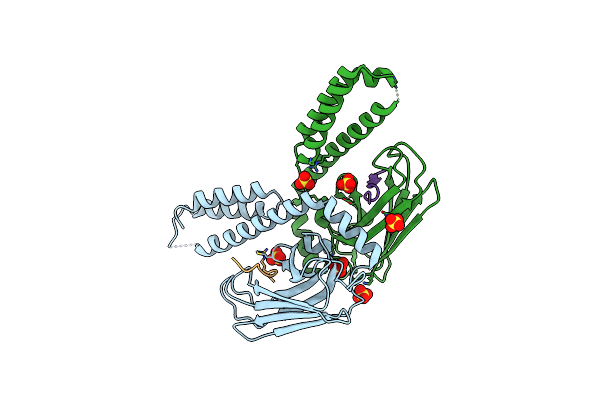
Crystal Structure Of The Substrate Binding Domain Of E.Coli Dnak In Complex With Sheep Bac7(1-21)
Organism: Escherichia coli, Ovis aries
Method: X-RAY DIFFRACTION Resolution:1.95 Å Release Date: 2013-11-13 Classification: CHAPERONE/Antibiotic Ligands: SO4 |
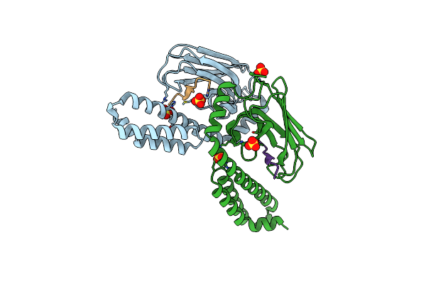 |
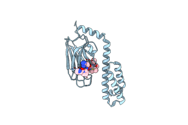
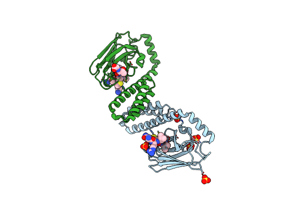
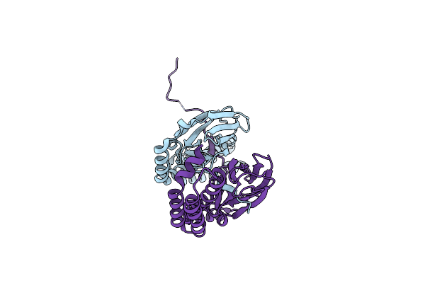
Crystal Structure Of The Substrate Binding Domain Of E.Coli Dnak In Complex With Sheep Bac7(35-43)
Organism: Escherichia coli, Ovis aries
Method: X-RAY DIFFRACTION Resolution:1.90 Å Release Date: 2013-11-13 Classification: CHAPERONE/ANTIBIOTIC Ligands: SO4 |
 |
Crystal Structure Of The Substrate Binding Domain Of E.Coli Dnak In Complex With Pyrrhocoricin
Organism: Escherichia coli, Pyrrhocoris apterus
Method: X-RAY DIFFRACTION Resolution:1.80 Å Release Date: 2013-04-17 Classification: CHAPERONE/PEPTIDE BINDING PROTEIN |
 |
Crystal Structure Of The Substrate Binding Domain Of E.Coli Dnak In Complex With Pr-39 (Residues 1 To 15)
Organism: Escherichia coli, Sus scrofa
Method: X-RAY DIFFRACTION Resolution:1.90 Å Release Date: 2013-04-17 Classification: CHAPERONE/PEPTIDE BINDING PROTEIN Ligands: SO4 |
 |
Crystal Structure Of The Substrate Binding Domain Of E.Coli Dnak In Complex With A3-Apo(Residues 1 To 20)
Organism: Escherichia coli
Method: X-RAY DIFFRACTION Resolution:1.65 Å Release Date: 2013-04-17 Classification: CHAPERONE/PEPTIDE BINDING PROTEIN Ligands: SO4 |
 |
Crystal Structure Of The Substrate Binding Domain Of E.Coli Dnak In Complex With The C-Terminal Part Of Pyrrhocoricin (Residues 12 To 20)
Organism: Escherichia coli, Pyrrhocoris apterus
Method: X-RAY DIFFRACTION Resolution:2.00 Å Release Date: 2013-04-17 Classification: CHAPERONE/PEPTIDE BINDING PROTEIN |
 |
Crystal Structure Of The Substrate Binding Domain Of E.Coli Dnak In Complex With The C-Terminal Part Of Drosocin (Residues 12 To 19)
Organism: Escherichia coli, Drosophila melanogaster
Method: X-RAY DIFFRACTION Resolution:1.90 Å Release Date: 2013-04-17 Classification: CHAPERONE/PEPTIDE BINDING PROTEIN |
 |
Crystal Structure Of The Substrate Binding Domain Of E.Coli Dnak In Complex With Heliocin (Residues 14 To 21)
Organism: Escherichia coli, Heliothis virescens
Method: X-RAY DIFFRACTION Resolution:2.00 Å Release Date: 2013-04-17 Classification: CHAPERONE/PEPTIDE BINDING PROTEIN |
 |
Crystal Structure Of The Substrate Binding Domain Of E.Coli Dnak In Complex With The Designer Peptide Nrllltg
Organism: Escherichia coli
Method: X-RAY DIFFRACTION Resolution:1.80 Å Release Date: 2013-04-17 Classification: CHAPERONE/PEPTIDE BINDING PROTEIN Ligands: SO4 |
 |
Crystal Structure Of The Substrate Binding Domain Of E.Coli Dnak In Complex With The Designer Peptide Nrlmltg
Organism: Escherichia coli
Method: X-RAY DIFFRACTION Resolution:1.70 Å Release Date: 2013-04-17 Classification: CHAPERONE/PEPTIDE BINDING PROTEIN Ligands: SO4 |
 |
Crystal Structure Of The Substrate Binding Domain Of E.Coli Dnak In Complex With The Designer Peptide Nrliltg
Organism: Escherichia coli
Method: X-RAY DIFFRACTION Resolution:1.85 Å Release Date: 2013-04-17 Classification: CHAPERONE Ligands: SO4 |
 |
Crystal Structure Of The Substrate Binding Domain Of E.Coli Dnak In Complex With The Designer Peptide Elplvki
Organism: Escherichia coli
Method: X-RAY DIFFRACTION Resolution:2.05 Å Release Date: 2013-04-17 Classification: CHAPERONE |
 |
Crystal Structure Of The Substrate Binding Domain Of E.Coli Dnak In Complex With An Apidaecin Fragment From The Bumblebee (Residues 3 To 11)
Organism: Escherichia coli, Bombus pascuorum
Method: X-RAY DIFFRACTION Resolution:1.95 Å Release Date: 2013-04-17 Classification: CHAPERONE/IMMUNE SYSTEM |
 |
Organism: Escherichia coli
Method: X-RAY DIFFRACTION Resolution:1.40 Å Release Date: 2013-04-17 Classification: CHAPERONE |
 |
Crystal Structure Of The Substrate Binding Domain Of E.Coli Dnak In Complex With Pyrrhocoricin_Lyzz (Residues 1 To 11)
Organism: Escherichia coli, Pyrrhocoris apterus
Method: X-RAY DIFFRACTION Resolution:1.55 Å Release Date: 2013-04-17 Classification: CHAPERONE, PEPTIDE BINDING PROTEIN Ligands: SO4 |
 |
Crystal Structure Of The Substrate Binding Domain Of E.Coli Dnak In Complex With Pyrrhocoricin_Lyzi (Residues 1 To 10)
Organism: Escherichia coli, Pyrrhocoris apterus
Method: X-RAY DIFFRACTION Resolution:1.70 Å Release Date: 2013-04-17 Classification: CHAPERONE, PEPTIDE BINDING PROTEIN Ligands: SO4 |
 |
Crystal Structure Of The Substrate Binding Domain Of E.Coli Dnak In Complex With A Short Apidaecin Peptide
Organism: Escherichia coli, Apis mellifera
Method: X-RAY DIFFRACTION Resolution:1.90 Å Release Date: 2012-05-30 Classification: CHAPERONE Ligands: SO4 |
 |
Crystal Structure Of The Substrate Binding Domain Of E.Coli Dnak In Complex With The Antimicrobial Peptide Oncocin
Organism: Escherichia coli, Oncopeltus fasciatus
Method: X-RAY DIFFRACTION Resolution:2.28 Å Release Date: 2011-03-23 Classification: CHAPERONE/ANTIMICROBIAL PROTEIN Ligands: SO4 |