
Search Count: 46
 |
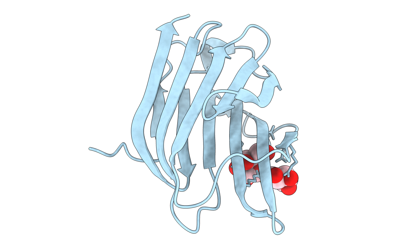
Crystal Structure Of Human Galectin-3 Crd In Complex With Diselenodigalactoside
Organism: Homo sapiens
Method: X-RAY DIFFRACTION Resolution:1.99 Å Release Date: 2022-07-13 Classification: SUGAR BINDING PROTEIN Ligands: 4IW, MG, CL |
 |
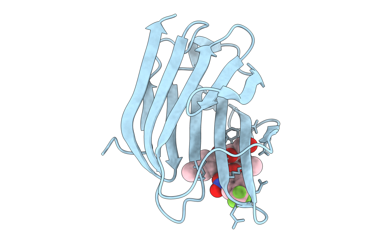
Crystal Structure Of Human Galectin-3 Crd In Complex With Selenodigalactoside
Organism: Homo sapiens
Method: X-RAY DIFFRACTION Resolution:1.96 Å Release Date: 2022-07-13 Classification: SUGAR BINDING PROTEIN Ligands: 4IZ, CL |
 |
Crystal Structure Of Human Galectin-3 Crd In Complex With Methyl 2-O-(2-Nitrobenzoyl)-3-O-(4-Methylbenzoyl)-B-D-Talopyranoside
Organism: Homo sapiens
Method: X-RAY DIFFRACTION Resolution:1.58 Å Release Date: 2022-07-13 Classification: SUGAR BINDING PROTEIN Ligands: CL, 57I |
 |
Crystal Structure Of Human Galectin-3 Crd In Complex With Methyl 2-O-(2-Nitro-4-Chloro)-Benzoyl-3-O-Toluoyl-B-D-Talopyranoside
Organism: Homo sapiens
Method: X-RAY DIFFRACTION Resolution:1.34 Å Release Date: 2022-07-13 Classification: SUGAR BINDING PROTEIN Ligands: CL, 59O |
 |
Crystal Structure Of Human Galectin-3 Crd In Complex With Methyl 2-O-(2-Nitro-4-Fluoro)-Benzoyl-3-O-Toluoyl-B-D-Talopyranoside
Organism: Homo sapiens
Method: X-RAY DIFFRACTION Resolution:1.49 Å Release Date: 2022-07-13 Classification: SUGAR BINDING PROTEIN Ligands: CL, 5A4 |
 |
Crystal Structure Of Human Galectin-3 Crd In Complex With Methyl 2-O-(2-Nitro-4-Trifluoromethyl-Benzoyl)-3-O-Toluoyl-B-D-Talopyranoside
Organism: Homo sapiens
Method: X-RAY DIFFRACTION Resolution:1.49 Å Release Date: 2022-07-13 Classification: SUGAR BINDING PROTEIN Ligands: CL, 5BI |
 |
Crystal Structure Of Human Galectin-3 Crd In Complex With Methyl 2-O-(3-Nitro-Benzoyl)-3-Toluoyl-B-D-Talopyranoside
Organism: Homo sapiens
Method: X-RAY DIFFRACTION Resolution:1.44 Å Release Date: 2022-07-13 Classification: SUGAR BINDING PROTEIN Ligands: CL, 5EI |
 |
Co-Crystallization Of Human Galectin-3 Crd Complex With Methyl 2-O-(2-Nitro-4-Chloro)-Benzoyl-3-O-Toluoyl-B-D-Talopyranoside
Organism: Homo sapiens
Method: X-RAY DIFFRACTION Resolution:1.05 Å Release Date: 2022-07-13 Classification: SUGAR BINDING PROTEIN Ligands: CL, 59O |
 |
Co-Crystallization Of Human Galectin-3 Crd Complex With Methyl 2-O-(2-Nitro-4-Fluoro)-Benzoyl-3-O-Toluoyl-B-D-Talopyranoside
Organism: Homo sapiens
Method: X-RAY DIFFRACTION Resolution:1.20 Å Release Date: 2022-07-13 Classification: SUGAR BINDING PROTEIN Ligands: CL, 5A4 |
 |
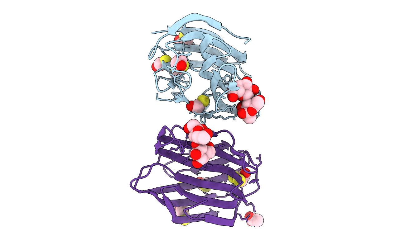
Crystal Structure Of Human Galectin-4 C-Terminal Carbohydrate Recognition Domain In Complex With Galactose Derivative
Organism: Homo sapiens
Method: X-RAY DIFFRACTION Resolution:2.28 Å Release Date: 2021-04-07 Classification: SUGAR BINDING PROTEIN Ligands: U61, GOL |
 |
Galectin-8N Terminal Domain In Complex With Methyl 3-O-[3-O-Benzyloxy]-Malonyl-Beta-D-Galactopyranoside
Organism: Homo sapiens
Method: X-RAY DIFFRACTION Resolution:1.59 Å Release Date: 2020-09-02 Classification: SUGAR BINDING PROTEIN Ligands: SZS, CL, EDO, PG4 |
 |
Crystal Structure Of Human Galectin-3 Crd In Complex With Methyl 3-O-(1-{3-O-[1-(B-D-Galactopyranosyl)-1,2,3-Triazol-4-Yl]-Methyl-B-D-Galactopyranosyl}-1,2,3-Triazol-4-Yl)-Methyl-B-D-Galactopyranoside
Organism: Homo sapiens
Method: X-RAY DIFFRACTION Resolution:1.99 Å Release Date: 2020-04-29 Classification: SUGAR BINDING PROTEIN Ligands: CL, P8G |
 |
Crystal Structure Of Human Galectin-3 Crd In Complex With Methyl 3-O-[1-(B-D-Galactopyranosyl)-1,2,3-Triazol-4-Yl]-Methyl-B-D-Galactopyranoside
Organism: Homo sapiens
Method: X-RAY DIFFRACTION Resolution:1.98 Å Release Date: 2020-04-29 Classification: SUGAR BINDING PROTEIN Ligands: P8J, CL |
 |
Organism: Homo sapiens
Method: X-RAY DIFFRACTION Resolution:1.28 Å Release Date: 2018-10-10 Classification: SUGAR BINDING PROTEIN Ligands: CL |
 |
Organism: Homo sapiens
Method: X-RAY DIFFRACTION Resolution:1.80 Å Release Date: 2018-10-10 Classification: SUGAR BINDING PROTEIN Ligands: BME, ACT |
 |
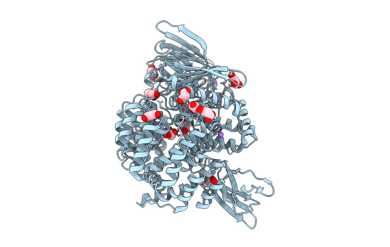
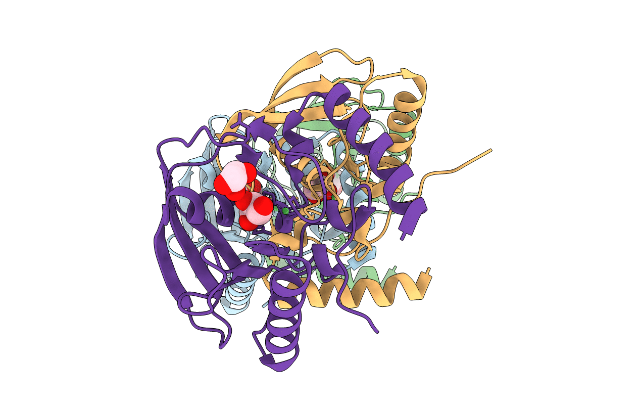
Crystal Structure Of Met260Ala Mutant Of E. Coli Aminopeptidase N In Complex With Bestatin
Organism: Escherichia coli (strain k12)
Method: X-RAY DIFFRACTION Resolution:2.30 Å Release Date: 2016-02-10 Classification: HYDROLASE Ligands: ZN, BES, NA, GOL, MLI |
 |
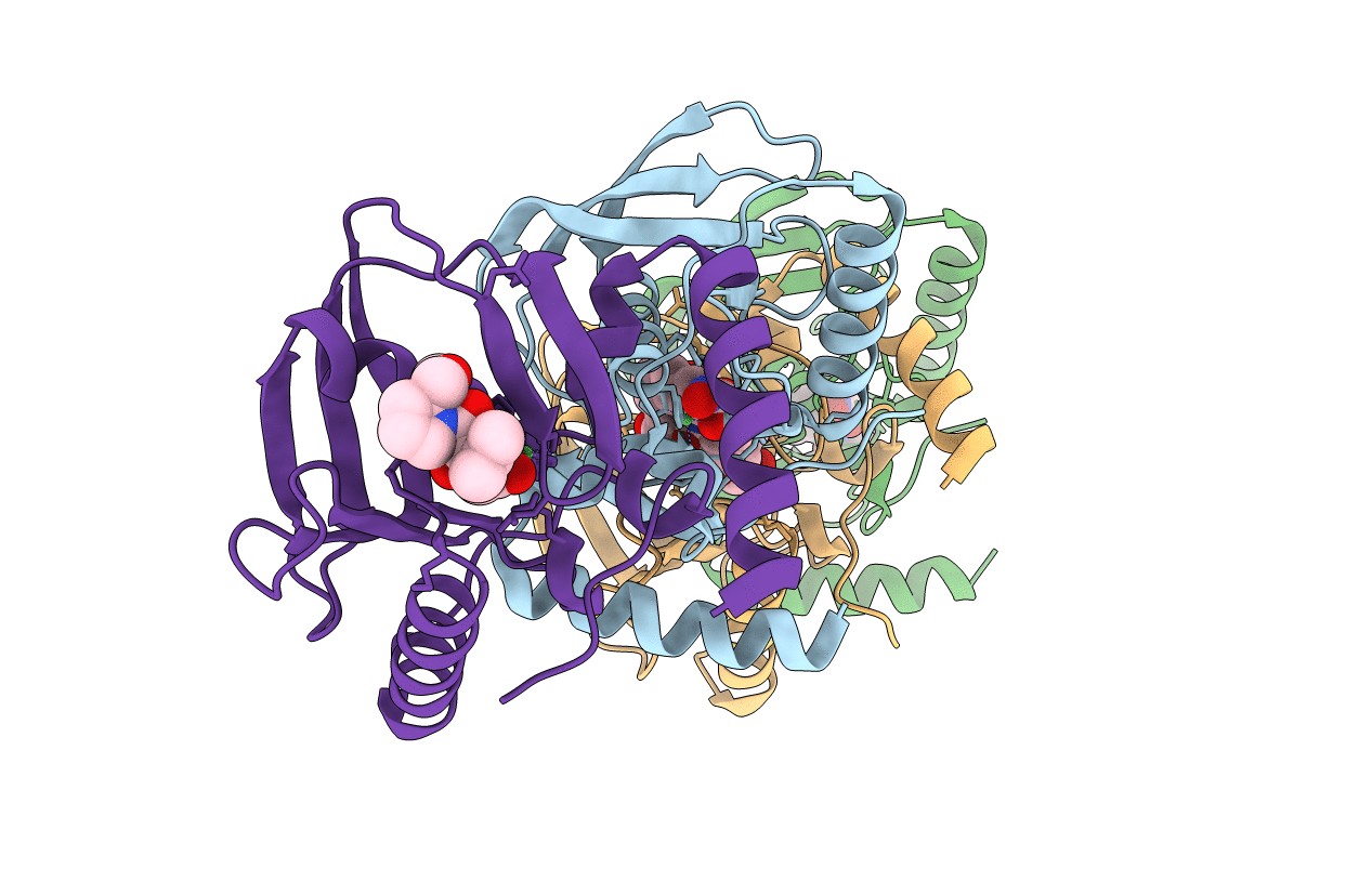
Crystal Structure Of Met260Ala Mutant Of E. Coli Aminopeptidase N In Complex With L-Phenylalanine
Organism: Escherichia coli (strain k12)
Method: X-RAY DIFFRACTION Resolution:2.66 Å Release Date: 2016-02-10 Classification: HYDROLASE Ligands: ZN, PHE, NA, GOL, MLI |
 |
Crystal Structure Of Dihydroorotate Dehydrogense From Mycobacterium Tuberculosis
Organism: Mycobacterium tuberculosis h37ra
Method: X-RAY DIFFRACTION Resolution:2.00 Å Release Date: 2016-01-20 Classification: OXIDOREDUCTASE Ligands: FMN, CL, MG |
 |
Organism: Haemophilus influenzae 86-028np
Method: X-RAY DIFFRACTION Resolution:2.05 Å Release Date: 2015-11-18 Classification: HYDROLASE Ligands: NI, GOL |
 |
Crystal Structure Of A Peptide Deformylase From Haemophilus Influenzae Complex With Actinonin
Organism: Haemophilus influenzae 86-028np
Method: X-RAY DIFFRACTION Resolution:2.33 Å Release Date: 2015-11-18 Classification: HYDROLASE Ligands: NI, BB2 |