
Search Count: 38
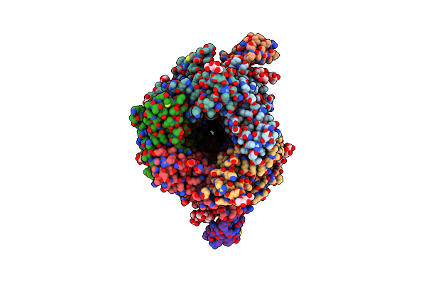 |
Organism: Laticauda semifasciata, Tetronarce californica
Method: ELECTRON MICROSCOPY Release Date: 2025-05-28 Classification: MEMBRANE PROTEIN Ligands: POV, NAG, OCT, PLM |
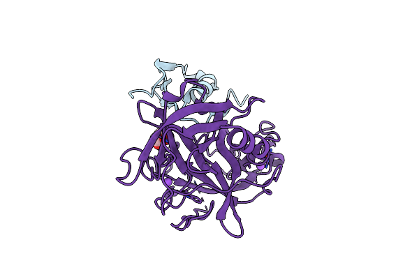 |
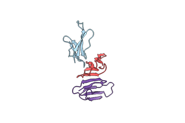
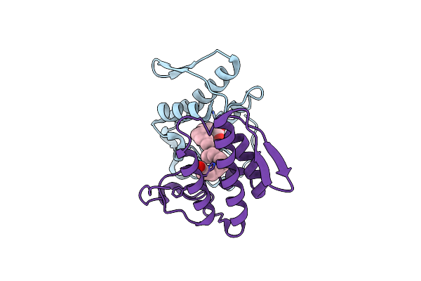
Crystal Structure Of Human Microplasmin In Complex With Kazal-Type Inhibitor Aati
Organism: Aedes aegypti, Homo sapiens
Method: X-RAY DIFFRACTION Resolution:1.95 Å Release Date: 2022-02-16 Classification: HYDROLASE Ligands: GOL, NA |
 |
Organism: Aedes aegypti
Method: X-RAY DIFFRACTION Resolution:2.08 Å Release Date: 2021-11-10 Classification: HYDROLASE Ligands: ZN |
 |
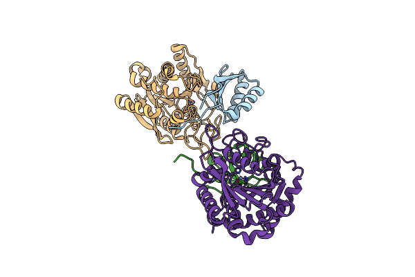
Crystal Structure Of Carboxypeptidase B Complexed With Potato Carboxypeptidase Inhibitor
Organism: Aedes aegypti, Solanum tuberosum
Method: X-RAY DIFFRACTION Resolution:2.20 Å Release Date: 2021-11-10 Classification: Hydrolase/Hydrolase inhibitor Ligands: ZN, GLY |
 |
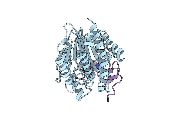
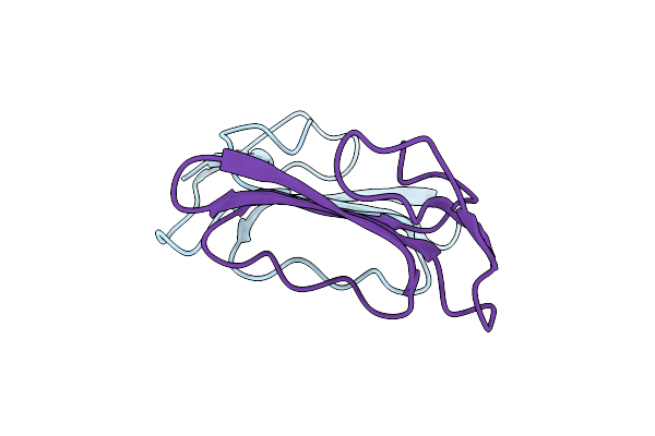
Unusual Quaternary Structure Of A Homodimeric Synergistic Toxin From Mamba Snake Venom
Organism: Dendroaspis jamesoni kaimosae
Method: X-RAY DIFFRACTION Resolution:2.40 Å Release Date: 2020-10-14 Classification: TOXIN Ligands: SO4 |
 |
Organism: Gallus gallus
Method: X-RAY DIFFRACTION Resolution:1.75 Å Release Date: 2019-03-13 Classification: HYDROLASE Ligands: CL, NA, GAI, GOL |
 |
Organism: Gallus gallus
Method: X-RAY DIFFRACTION Resolution:1.75 Å Release Date: 2019-03-13 Classification: HYDROLASE Ligands: GAI, CL, GOL |
 |
Organism: Gallus gallus
Method: X-RAY DIFFRACTION Resolution:1.76 Å Release Date: 2019-03-13 Classification: HYDROLASE Ligands: GAI, NA, CL |
 |
Organism: Gallus gallus
Method: X-RAY DIFFRACTION Resolution:1.75 Å Release Date: 2019-03-13 Classification: HYDROLASE Ligands: CL, NA, GAI, GOL |
 |
Hen Egg White Lysozyme Native Crystals Soaked For 2 Hours In Precipitant Solution Containing 1 M Guanidine Hydrochloride And 25% Glycerol, Before Data Collection
Organism: Gallus gallus
Method: X-RAY DIFFRACTION Resolution:2.00 Å Release Date: 2017-09-13 Classification: HYDROLASE Ligands: CL, GAI |
 |
Organism: Homo sapiens, Amblyomma variegatum
Method: X-RAY DIFFRACTION Resolution:2.09 Å Release Date: 2017-05-03 Classification: hydrolase/hydrolase inhibitor |
 |
Organism: Micrurus fulvius
Method: X-RAY DIFFRACTION Resolution:1.95 Å Release Date: 2016-05-18 Classification: TOXIN Ligands: ZN |
 |
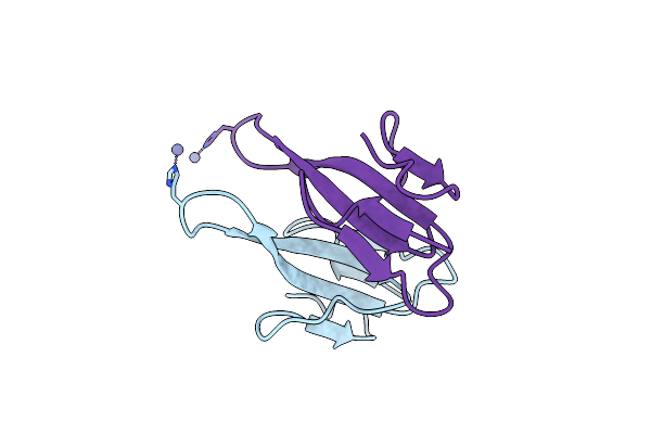
Ringhalexin From Hemachatus Haemachatus: A Novel Inhibitor Of Extrinsic Tenase Complex
Organism: Hemachatus haemachatus
Method: X-RAY DIFFRACTION Resolution:2.95 Å Release Date: 2016-05-11 Classification: TOXIN |
 |
Organism: Hemachatus haemachatus
Method: X-RAY DIFFRACTION Resolution:2.43 Å Release Date: 2012-11-07 Classification: TOXIN |
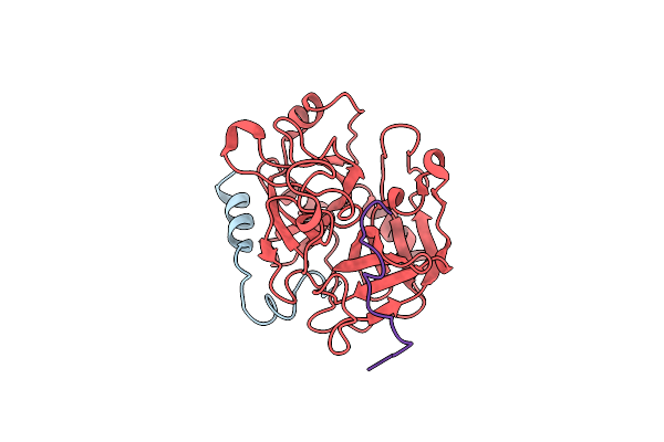 |
Crystal Structure Of Thrombin-Variegin Complex: Insights Of A Novel Mechanism Of Inhibition And Design Of Tunable Thrombin Inhibitors
Organism: Homo sapiens, Amblyomma variegatum
Method: X-RAY DIFFRACTION Resolution:2.40 Å Release Date: 2011-11-23 Classification: HYDROLASE/HYDROLASE INHIBITOR |
 |
Crystal Structure Of Beta-Cardiotoxin, A Novel Three-Finger Cardiotoxin From The Venom Of Ophiophagus Hannah
Organism: Ophiophagus hannah
Method: X-RAY DIFFRACTION Resolution:2.41 Å Release Date: 2011-11-16 Classification: TOXIN |
 |
Structural And Functional Characterization Of A Novel Homodimeric Three-Finger Neurotoxin From The Venom Of Ophiophagus Hannah (King Cobra)
Organism: Ophiophagus hannah
Method: X-RAY DIFFRACTION Resolution:1.55 Å Release Date: 2010-01-12 Classification: TOXIN |
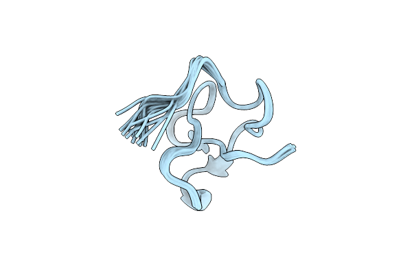 |
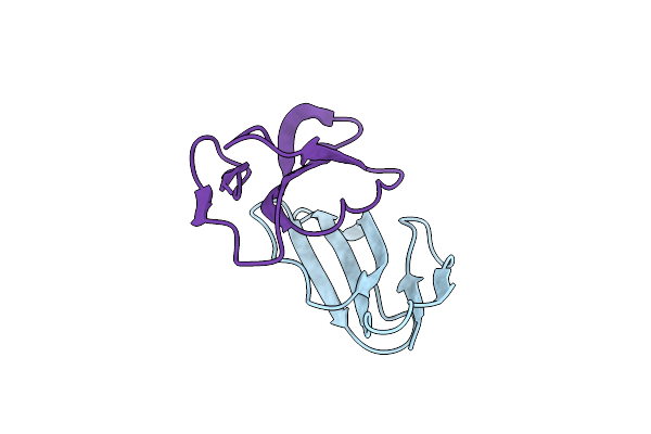
Antibacterial Peptide From Eggshell Matrix: Structure And Self-Assembly Of Beta-Defensin Like Peptide From The Chinese Soft-Shelled Turtle Eggshell
Organism: Pelodiscus sinensis
Method: SOLUTION NMR Release Date: 2008-05-27 Classification: ANTIMICROBIAL PROTEIN |
 |
Organism: Echis carinatus
Method: X-RAY DIFFRACTION Resolution:2.30 Å Release Date: 2007-12-18 Classification: HYDROLASE Ligands: SVR |
 |
Organism: Echis carinatus
Method: X-RAY DIFFRACTION Resolution:1.95 Å Release Date: 2007-10-16 Classification: HYDROLASE Ligands: DAO |