
Search Count: 42
 |
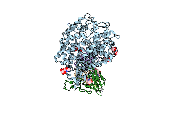
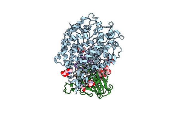
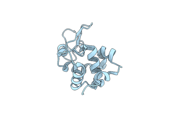
Organism: Homo sapiens, Oryctolagus cuniculus
Method: ELECTRON MICROSCOPY Release Date: 2025-08-13 Classification: CYTOSOLIC PROTEIN/CONTRACTILE PROTEIN |
 |
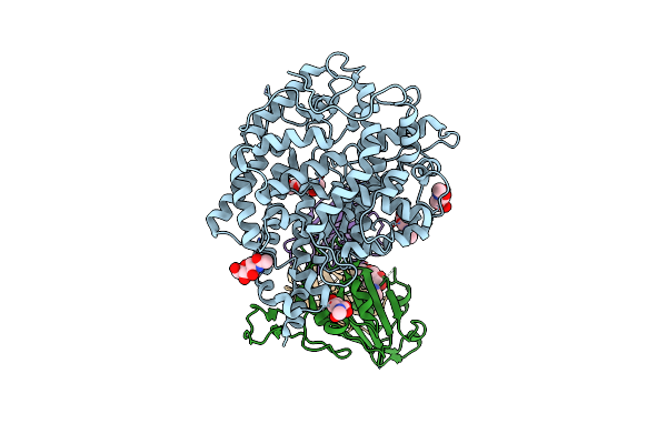
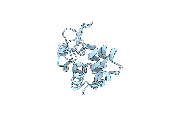
Organism: Homo sapiens, Oryctolagus cuniculus
Method: ELECTRON MICROSCOPY Release Date: 2025-08-13 Classification: CYTOSOLIC PROTEIN/CONTRACTILE PROTEIN |
 |
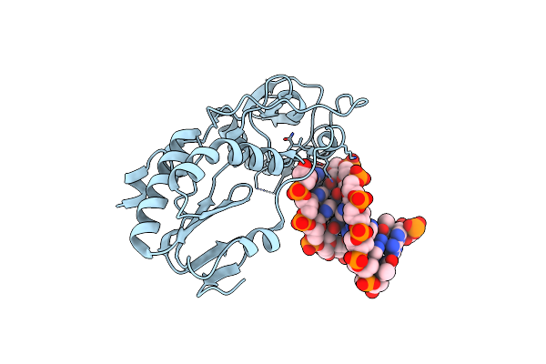
Sars-Cov-2 Xbb.1 Spike Rbd Bound To The Human Ace2 Ectodomain And The S309 Neutralizing Antibody Fab Fragment
Organism: Homo sapiens, Severe acute respiratory syndrome coronavirus
Method: ELECTRON MICROSCOPY Release Date: 2023-10-04 Classification: VIRAL PROTEIN/IMMUNE SYSTEM Ligands: NAG |
 |
Sars-Cov-2 Bq.1.1 Spike Rbd Bound To The Human Ace2 Ectodomain And The S309 Neutralizing Antibody Fab Fragment
Organism: Homo sapiens, Severe acute respiratory syndrome coronavirus 2
Method: ELECTRON MICROSCOPY Release Date: 2023-10-04 Classification: VIRAL PROTEIN/IMMUNE SYSTEM Ligands: NAG, ZN |
 |
Sars-Cov-2 Bn.1 Spike Rbd Bound To The Human Ace2 Ectodomain And The S309 Neutralizing Antibody Fab Fragment
Organism: Homo sapiens, Severe acute respiratory syndrome coronavirus
Method: ELECTRON MICROSCOPY Release Date: 2023-10-04 Classification: VIRAL PROTEIN Ligands: NAG |
 |
Organism: Rattus norvegicus, Gallus gallus, Bos taurus
Method: X-RAY DIFFRACTION Resolution:2.10 Å Release Date: 2022-12-28 Classification: CELL CYCLE Ligands: GTP, MG, CA, GDP, MES, GOL, SOZ, EDO, IMD, ACP |
 |
Organism: Gallus gallus
Method: X-RAY DIFFRACTION Resolution:1.60 Å Release Date: 2021-03-31 Classification: HYDROLASE |
 |
Organism: Gallus gallus
Method: X-RAY DIFFRACTION Resolution:1.60 Å Release Date: 2021-03-31 Classification: HYDROLASE |
 |
Organism: Gallus gallus
Method: X-RAY DIFFRACTION Resolution:1.65 Å Release Date: 2021-03-31 Classification: HYDROLASE |
 |
Organism: Gallus gallus
Method: X-RAY DIFFRACTION Resolution:1.65 Å Release Date: 2021-03-31 Classification: HYDROLASE |
 |
Organism: Gallus gallus
Method: X-RAY DIFFRACTION Resolution:1.70 Å Release Date: 2021-03-31 Classification: HYDROLASE |
 |
Organism: Gallus gallus
Method: X-RAY DIFFRACTION Resolution:1.70 Å Release Date: 2021-03-31 Classification: HYDROLASE |
 |
Organism: Micrococcus luteus
Method: X-RAY DIFFRACTION Resolution:2.25 Å Release Date: 2020-08-19 Classification: OXIDOREDUCTASE |
 |
Organism: Micrococcus luteus
Method: X-RAY DIFFRACTION Resolution:2.26 Å Release Date: 2020-08-19 Classification: OXIDOREDUCTASE |
 |
Organism: Moloney murine leukemia virus (isolate shinnick), Synthetic construct
Method: X-RAY DIFFRACTION Resolution:1.70 Å Release Date: 2019-02-27 Classification: HYDROLASE/DNA |
 |
Organism: Moloney murine leukemia virus (isolate shinnick), Synthetic construct
Method: X-RAY DIFFRACTION Resolution:1.60 Å Release Date: 2019-02-27 Classification: HYDROLASE/DNA |
 |
Organism: Moloney murine leukemia virus (isolate shinnick), Synthetic construct
Method: X-RAY DIFFRACTION Resolution:1.70 Å Release Date: 2019-02-27 Classification: HYDROLASE/DNA |
 |
Hydrogen Bonding Complementary, Not Size Complementarity Is Key In The Formation Of The Double Helix
Organism: Moloney murine leukemia virus, Escherichia coli
Method: X-RAY DIFFRACTION Resolution:1.90 Å Release Date: 2018-09-19 Classification: DNA BINDING PROTEIN/DNA |
 |
Hydrogen Bonding Complementary, Not Size Complementarity Is Key In The Formation Of The Double Helix
Organism: Moloney murine leukemia virus, Synthetic construct
Method: X-RAY DIFFRACTION Resolution:1.69 Å Release Date: 2018-09-19 Classification: DNA BINDING PROTEIN/DNA |
 |
Hydrogen Bonding Complementary, Not Size Complementarity Is Key In The Formation Of The Double Helix
Organism: Moloney murine leukemia virus, Escherichia coli
Method: X-RAY DIFFRACTION Resolution:2.00 Å Release Date: 2018-09-19 Classification: DNA BINDING PROTEIN/DNA |