
Search Count: 33
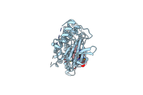 |
Design, Synthesis, Biological Evaluation, And X-Ray Crystallography Of Diarylpyrazole Derivatives Possessing Terminal Arylsulfonamide Moieties As Anti-Proliferative Agents Targeting C-Jun N-Terminal Kinase (Jnk)
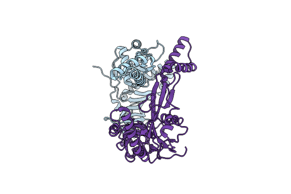
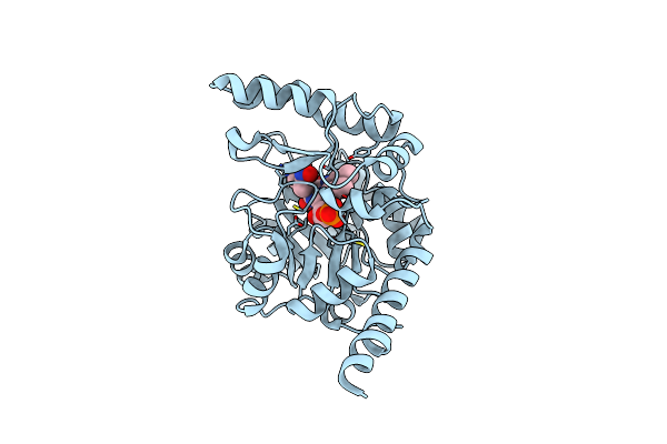
Organism: Homo sapiens
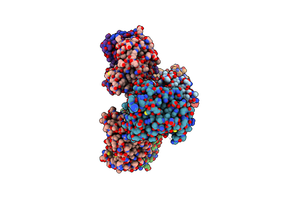
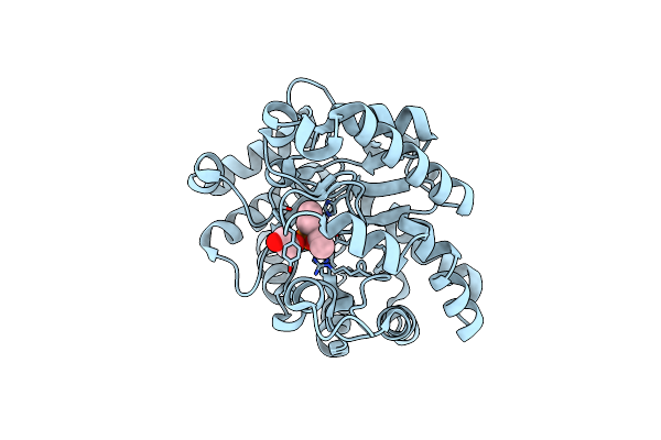
Method: X-RAY DIFFRACTION Resolution:2.81 Å Release Date: 2023-10-18 Classification: HYDROLASE Ligands: WNK |
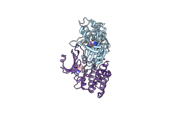 |
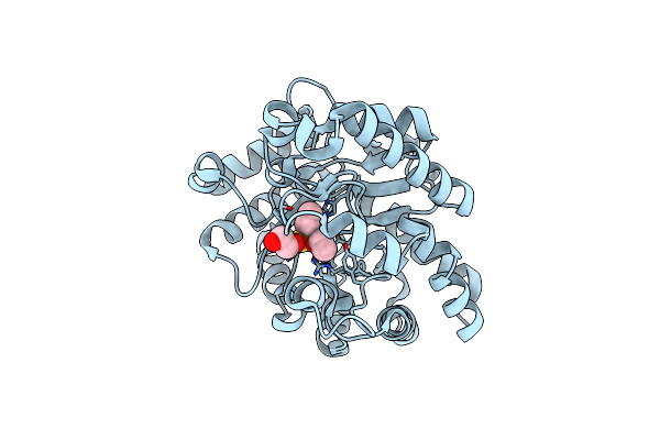
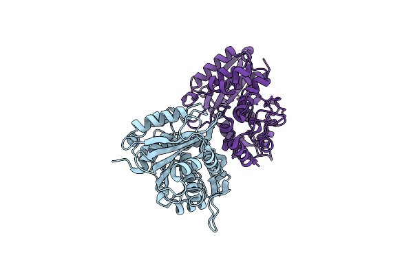
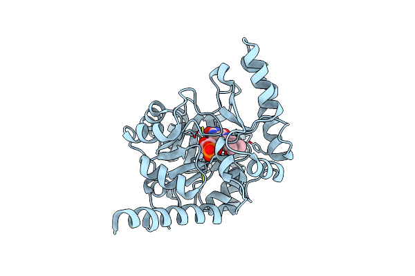
Organism: Homo sapiens
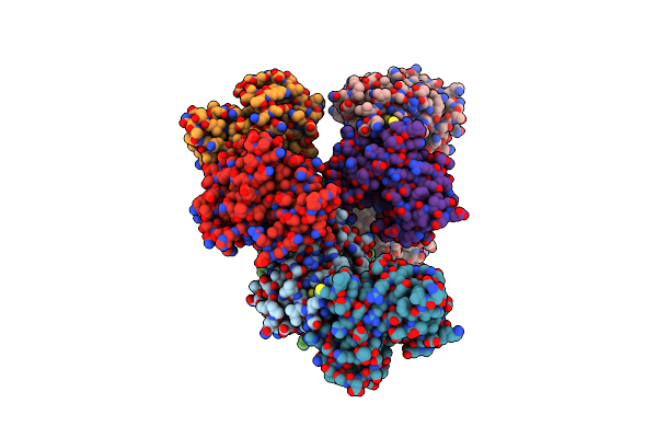
Method: X-RAY DIFFRACTION Resolution:2.10 Å Release Date: 2021-08-18 Classification: SIGNALING PROTEIN Ligands: RXT |
 |
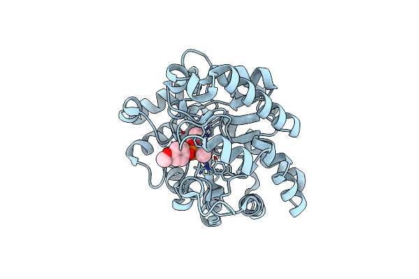
Organism: Homo sapiens
Method: X-RAY DIFFRACTION Resolution:2.15 Å Release Date: 2019-10-02 Classification: CHAPERONE |
 |
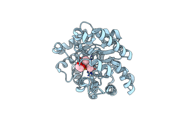
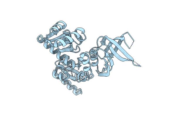
Organism: Homo sapiens
Method: X-RAY DIFFRACTION Resolution:1.40 Å Release Date: 2019-10-02 Classification: CHAPERONE |
 |
Organism: Homo sapiens
Method: X-RAY DIFFRACTION Resolution:1.90 Å Release Date: 2018-11-21 Classification: RNA BINDING PROTEIN/LIGASE |
 |
Organism: Homo sapiens
Method: X-RAY DIFFRACTION Resolution:2.60 Å Release Date: 2015-10-21 Classification: LIGASE Ligands: GOL |
 |
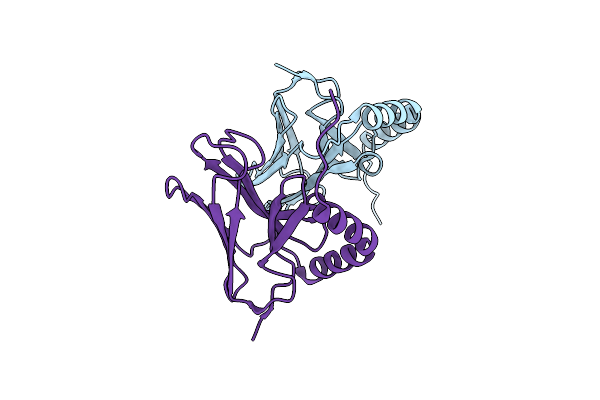
The Crystal Structure Of The Gst-Like Domains Complex Of Aimp3-Eprs Mutant C92Sc105Sc123S
Organism: Homo sapiens
Method: X-RAY DIFFRACTION Resolution:2.60 Å Release Date: 2015-10-21 Classification: TRANSLATION/LIGASE |
 |
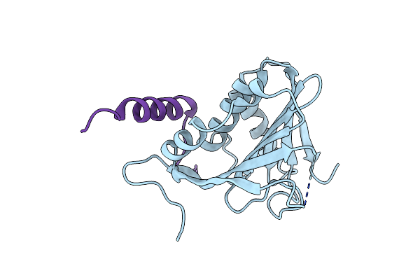
Organism: Homo sapiens
Method: X-RAY DIFFRACTION Resolution:1.60 Å Release Date: 2014-07-16 Classification: LIGASE Ligands: I3C |
 |
Opda From Agrobacterium Radiobacter With Bound Product Diethyl Thiophosphate From Crystal Soaking With Tetraethyl Dithiopyrophosphate- 1.8 A
Organism: Agrobacterium tumefaciens
Method: X-RAY DIFFRACTION Resolution:1.80 Å Release Date: 2008-02-19 Classification: HYDROLASE Ligands: FE2, CO, DPJ, EDO |
 |
Opda From Agrobacterium Radiobacter With Bound Diethyl Phosphate From Crystal Soaking With The Compound- 1.9 A
Organism: Agrobacterium tumefaciens
Method: X-RAY DIFFRACTION Resolution:2.10 Å Release Date: 2008-02-12 Classification: HYDROLASE Ligands: FE2, CO, DPF, EDO |
 |
Opda From Agrobacterium Radiobacter With Bound Diethyl Thiophosphate From Crystal Soaking With The Compound- 1.95 A
Organism: Agrobacterium tumefaciens
Method: X-RAY DIFFRACTION Resolution:1.95 Å Release Date: 2008-02-12 Classification: HYDROLASE Ligands: FE2, CO, DPJ, EDO |
 |
Opda From Agrobacterium Radiobacter With Bound Product Diethyl Phosphate From Crystal Soaking With Diethyl 4-Methoxyphenyl Phosphate (450H)- 2.5 A
Organism: Agrobacterium tumefaciens
Method: X-RAY DIFFRACTION Resolution:2.50 Å Release Date: 2008-02-12 Classification: HYDROLASE Ligands: FE2, CO, DPF, EDO |
 |
Opda From Agrobacterium Radiobacter With Bound Slow Substrate Diethyl 4-Methoxyphenyl Phosphate (20H)- 1.7 A
Organism: Agrobacterium tumefaciens
Method: X-RAY DIFFRACTION Resolution:1.70 Å Release Date: 2008-02-12 Classification: HYDROLASE Ligands: FE2, CO, EPL |
 |
Opda From Agrobacterium Radiobacter With Bound Product Diethyl Thiophosphate From Co-Crystallisation With Tetraethyl Dithiopyrophosphate- 1.8 A
Organism: Agrobacterium tumefaciens
Method: X-RAY DIFFRACTION Resolution:1.80 Å Release Date: 2008-02-12 Classification: HYDROLASE Ligands: FE2, CO, DPJ, EDO |
 |
Organism: Photobacterium sp. m37
Method: X-RAY DIFFRACTION Resolution:2.20 Å Release Date: 2008-01-29 Classification: HYDROLASE |
 |
The Recombination-Associated Protein Rdgc Adopts A Novel Toroidal Architecture For Dna Binding
Organism: Pseudomonas aeruginosa
Method: X-RAY DIFFRACTION Resolution:2.50 Å Release Date: 2007-11-20 Classification: DNA BINDING PROTEIN |
 |
Organism: Thermoplasma acidophilum
Method: X-RAY DIFFRACTION Resolution:2.90 Å Release Date: 2007-10-09 Classification: CELL CYCLE |
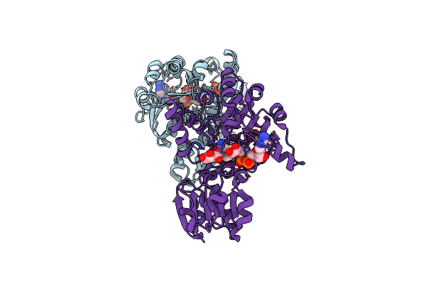 |
Crystal Structure Of D-Erythronate-4-Phosphate Dehydrogenase Complexed With Nad
Organism: Pseudomonas aeruginosa
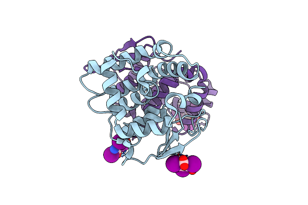
Method: X-RAY DIFFRACTION Resolution:2.30 Å Release Date: 2007-02-20 Classification: OXIDOREDUCTASE Ligands: PO4, NAD, TLA, GOL |
 |
Organism: Pseudomonas aeruginosa pao1
Method: X-RAY DIFFRACTION Resolution:2.00 Å Release Date: 2006-05-16 Classification: OXIDOREDUCTASE Ligands: FMN |
 |
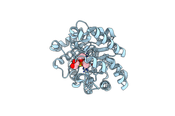
Crystal Structure Of 2-Nitropropane Dioxygenase Complexed With Fmn And Substrate
Organism: Pseudomonas aeruginosa pao1
Method: X-RAY DIFFRACTION Resolution:2.30 Å Release Date: 2006-05-16 Classification: OXIDOREDUCTASE Ligands: FMN, NIS |