
Search Count: 17
 |
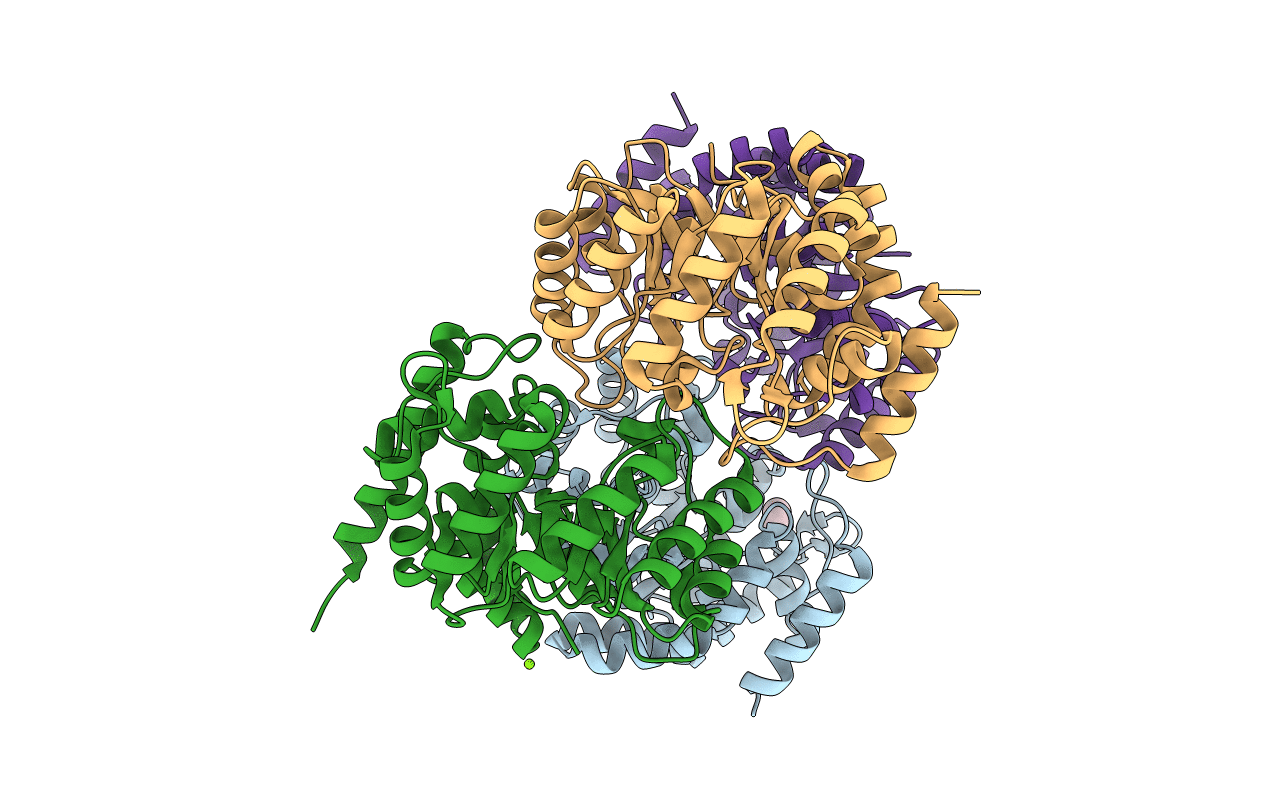
Organism: Homo sapiens
Method: X-RAY DIFFRACTION Release Date: 2025-07-16 Classification: METAL BINDING PROTEIN Ligands: ZN |
 |
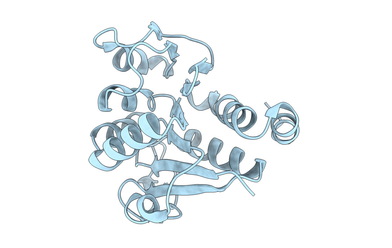
Organism: Homo sapiens
Method: X-RAY DIFFRACTION Release Date: 2025-07-16 Classification: METAL BINDING PROTEIN, LYASE Ligands: ZN |
 |
Organism: Homo sapiens
Method: X-RAY DIFFRACTION Release Date: 2025-07-16 Classification: METAL BINDING PROTEIN, LYASE Ligands: ZN, CO2 |
 |
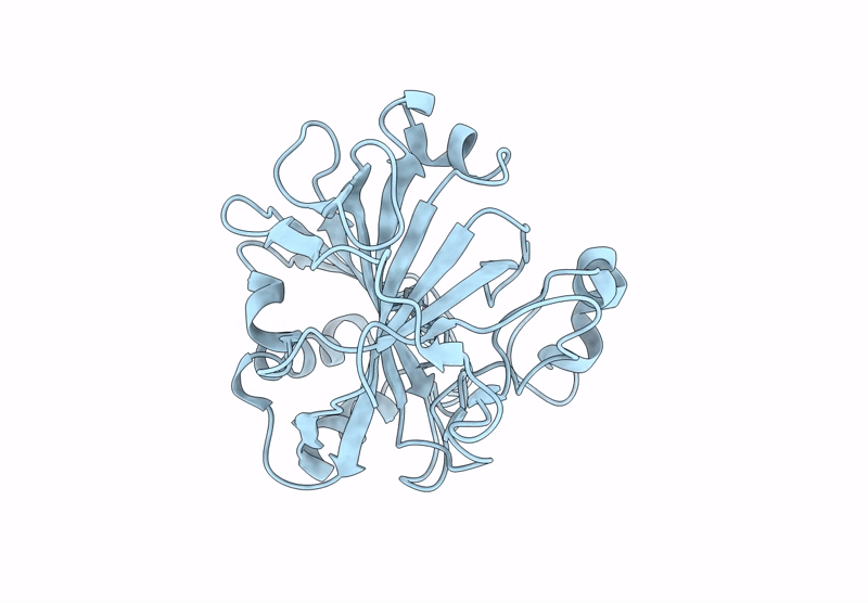
Organism: Homo sapiens
Method: X-RAY DIFFRACTION Release Date: 2025-07-16 Classification: LYASE Ligands: ZN, BCT |
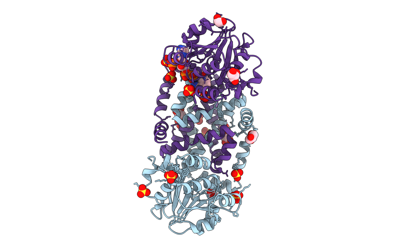 |
Organism: Streptococcus pneumoniae d39
Method: X-RAY DIFFRACTION Resolution:1.69 Å Release Date: 2020-08-12 Classification: ISOMERASE Ligands: GOL, SO4, NAP, MG |
 |
Organism: Streptococcus pneumoniae d39
Method: X-RAY DIFFRACTION Resolution:1.95 Å Release Date: 2020-08-12 Classification: ISOMERASE Ligands: SO4, GOL |
 |
Organism: Streptococcus pneumoniae d39
Method: X-RAY DIFFRACTION Resolution:2.02 Å Release Date: 2020-08-12 Classification: ISOMERASE |
 |
Organism: Streptococcus pneumoniae d39
Method: X-RAY DIFFRACTION Resolution:2.29 Å Release Date: 2020-08-12 Classification: ISOMERASE |
 |
Organism: Streptococcus pneumoniae d39
Method: X-RAY DIFFRACTION Resolution:2.02 Å Release Date: 2020-08-12 Classification: ISOMERASE Ligands: NAP, SO4, GOL, NH4 |
 |
Two Intermediate States Of Conformation Switch In Dual Specificity Phosphatase 13A
Organism: Homo sapiens
Method: X-RAY DIFFRACTION Resolution:1.69 Å Release Date: 2018-04-11 Classification: HYDROLASE Ligands: PO4 |
 |
Organism: Clonorchis sinensis
Method: X-RAY DIFFRACTION Resolution:1.90 Å Release Date: 2010-09-08 Classification: TRANSFERASE Ligands: GSH, ZN, SO4 |
 |
Crystal Structure Of Escherichia Coli Mazg, The Regulator Of Nutritional Stress Response
Organism: Escherichia coli
Method: X-RAY DIFFRACTION Resolution:2.10 Å Release Date: 2008-04-22 Classification: HYDROLASE |
 |
Crystal Structure Of Escherichia Coli Mazg, The Regulator Of Nutritional Stress Response
Organism: Escherichia coli
Method: X-RAY DIFFRACTION Release Date: 2008-04-22 Classification: HYDROLASE Ligands: MG, ATP |
 |
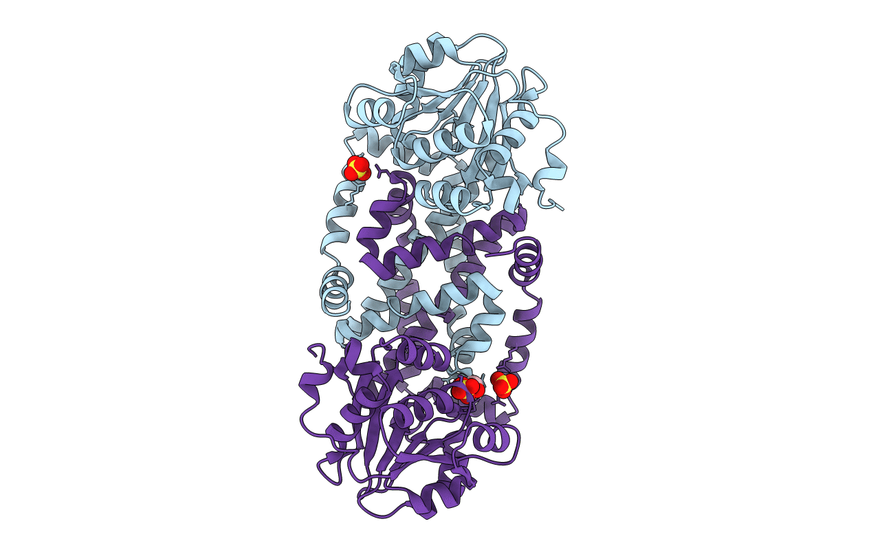
Crystal Structure Of Dihydrodipicolinate Synthase From Hahella Chejuensis At 1.5A Resolution
Organism: Hahella chejuensis
Method: X-RAY DIFFRACTION Resolution:1.50 Å Release Date: 2007-10-23 Classification: LYASE Ligands: MG, EOH |
 |
Organism: Homo sapiens
Method: X-RAY DIFFRACTION Resolution:2.50 Å Release Date: 2004-02-03 Classification: RNA BINDING PROTEIN |
 |
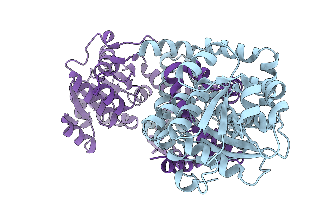
Organism: Escherichia coli
Method: X-RAY DIFFRACTION Resolution:2.80 Å Release Date: 2003-10-16 Classification: chaperone,hydrolase |
 |
Organism: Escherichia coli
Method: X-RAY DIFFRACTION Resolution:2.31 Å Release Date: 2003-10-16 Classification: chaperone,hydrolase |