
Search Count: 6
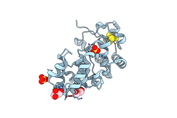 |
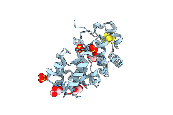
Crystal Structure Of The Core Fragment Of Muty From E.Coli At 1.2A Resolution
Organism: Escherichia coli
Method: X-RAY DIFFRACTION Resolution:1.20 Å Release Date: 2002-11-26 Classification: HYDROLASE Ligands: SO4, SF4, GOL |
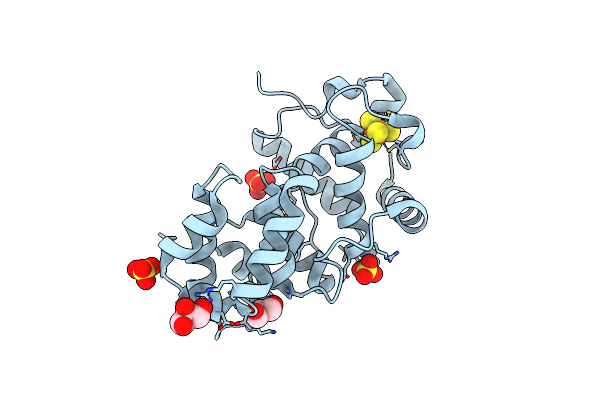 |
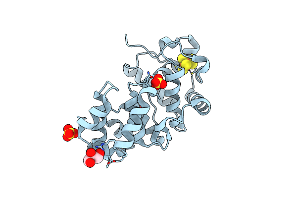
Crystal Structure Of The Core Fragment Of Muty From E.Coli At 1.55A Resolution
Organism: Escherichia coli
Method: X-RAY DIFFRACTION Resolution:1.55 Å Release Date: 2002-11-26 Classification: HYDROLASE Ligands: SO4, SF4, GOL |
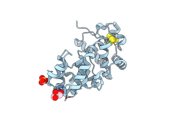 |
Organism: Escherichia coli
Method: X-RAY DIFFRACTION Resolution:1.60 Å Release Date: 2002-11-26 Classification: HYDROLASE Ligands: SO4, SF4, GOL |
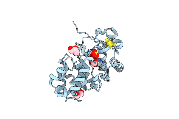 |
Organism: Escherichia coli
Method: X-RAY DIFFRACTION Resolution:1.35 Å Release Date: 2002-11-26 Classification: HYDROLASE Ligands: SO4, SF4, GOL |
 |
Organism: Escherichia coli
Method: X-RAY DIFFRACTION Resolution:1.50 Å Release Date: 2002-11-26 Classification: HYDROLASE Ligands: SO4, SF4, GOL |
 |
Organism: Escherichia coli
Method: X-RAY DIFFRACTION Resolution:1.50 Å Release Date: 2002-11-26 Classification: HYDROLASE Ligands: SO4, SF4, GOL |