
Search Count: 37
 |
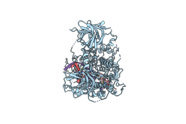
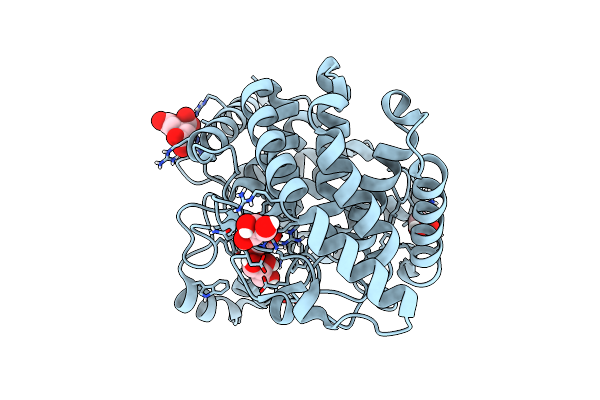
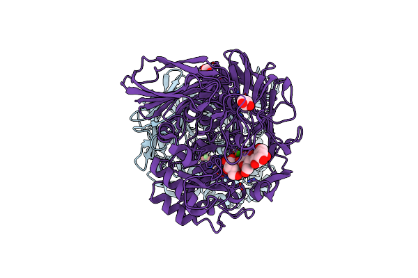
Cryo-Em Structure Of Arabidopsis Thaliana Met1 (Aa:621-1534) In Complex With Hemimethylated Dna Analog
Organism: Arabidopsis thaliana, Synthetic construct
Method: ELECTRON MICROSCOPY Release Date: 2025-08-20 Classification: STRUCTURAL PROTEIN/DNA Ligands: SAH, ZN |
 |
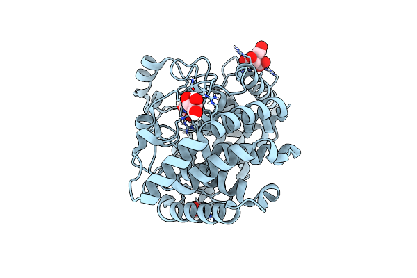
Organism: Arabidopsis thaliana
Method: ELECTRON MICROSCOPY Release Date: 2025-08-20 Classification: STRUCTURAL PROTEIN Ligands: ZN |
 |
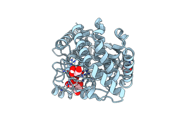
Organism: Homo sapiens
Method: ELECTRON MICROSCOPY Release Date: 2025-06-25 Classification: STRUCTURAL PROTEIN Ligands: ZN |
 |
Organism: Homo sapiens
Method: ELECTRON MICROSCOPY Release Date: 2025-06-25 Classification: STRUCTURAL PROTEIN |
 |
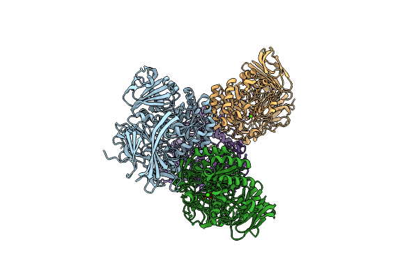
Cryo-Em Structure Of Human Dnmt1 (Aa:351-1616) In Complex With Ubiquitinated Paf15 And Hemimethylated Dna Analog
Organism: Homo sapiens, Synthetic construct
Method: ELECTRON MICROSCOPY Release Date: 2025-01-15 Classification: TRANSFERASE Ligands: SAH, ZN |
 |
Cryo-Em Structure Of Cdca7 Bound To Nucleosome Including Hemimethylated Cpg Site In Widom601 Positioning Sequence.
Organism: Homo sapiens, Synthetic construct
Method: ELECTRON MICROSCOPY Release Date: 2024-07-31 Classification: DNA BINDING PROTEIN Ligands: ZN |
 |
Cryo-Em Structure Of Human Dnmt1 (Aa:351-1616) In Complex With Ubiquitinated H3 And Hemimethylated Dna Analog (Cxxc-Ordered Form)
Organism: Homo sapiens, Synthetic construct
Method: ELECTRON MICROSCOPY Release Date: 2022-11-30 Classification: TRANSFERASE/DNA Ligands: ZN, SAH |
 |
Cryo-Em Structure Of Human Dnmt1 (Aa:351-1616) In Complex With Ubiquitinated H3 And Hemimethylated Dna Analog (Cxxc-Disordered Form)
Organism: Homo sapiens, Synthetic construct
Method: ELECTRON MICROSCOPY Release Date: 2022-11-30 Classification: TRANSFERASE/DNA Ligands: ZN |
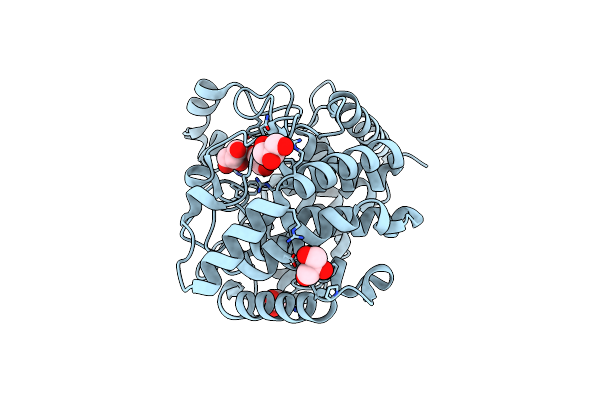 |
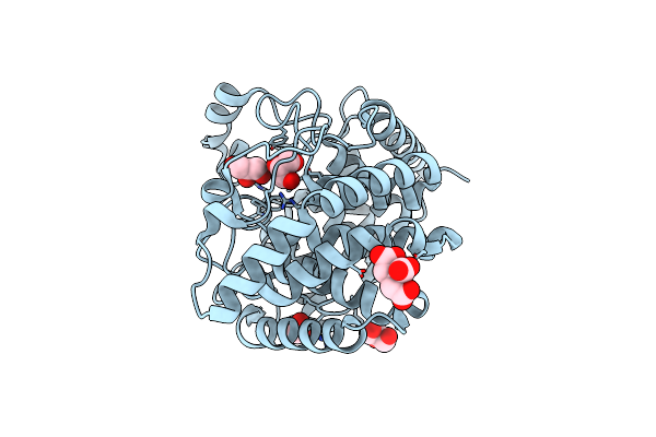
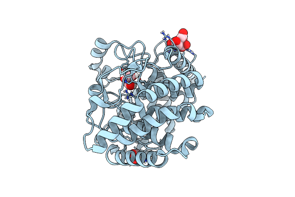
Organism: Kribbella flavida (strain dsm 17836 / jcm 10339 / nbrc 14399)
Method: X-RAY DIFFRACTION Resolution:1.71 Å Release Date: 2021-05-12 Classification: HYDROLASE Ligands: GOL, NH4 |
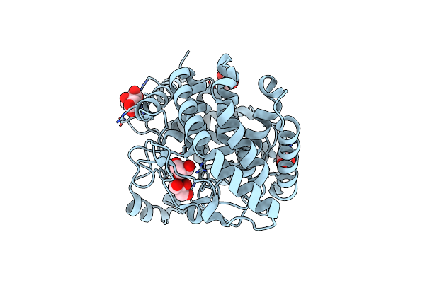 |
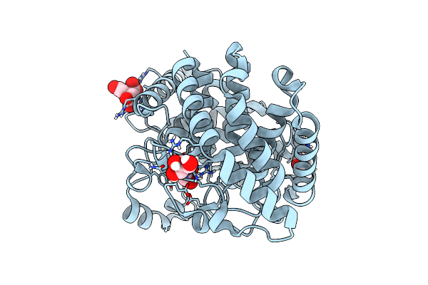
Organism: Kribbella flavida (strain dsm 17836 / jcm 10339 / nbrc 14399)
Method: X-RAY DIFFRACTION Resolution:1.51 Å Release Date: 2021-05-12 Classification: HYDROLASE Ligands: GOL, CIT, NH4 |
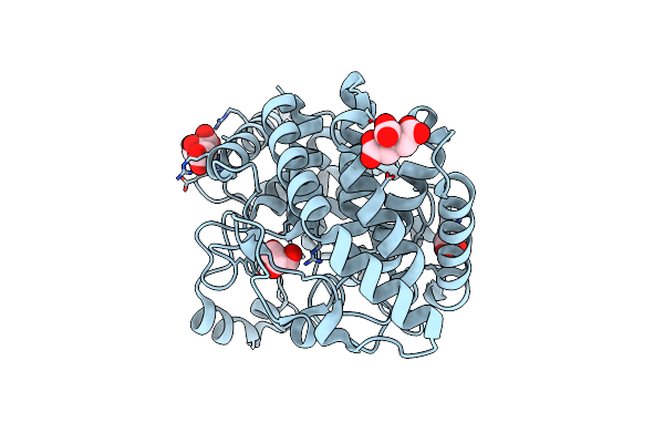 |
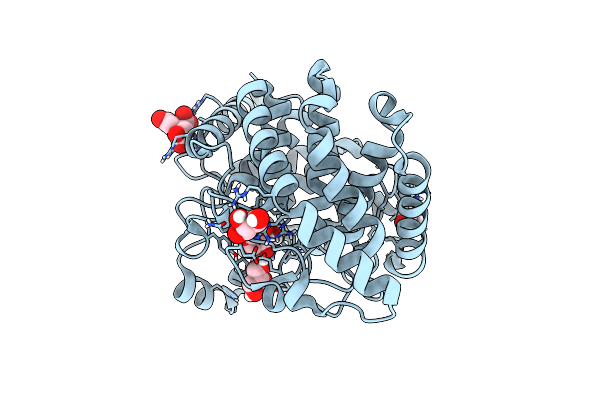
Organism: Kribbella flavida (strain dsm 17836 / jcm 10339 / nbrc 14399)
Method: X-RAY DIFFRACTION Resolution:1.80 Å Release Date: 2021-05-12 Classification: HYDROLASE Ligands: GOL, CIT, NH4 |
 |
Organism: Kribbella flavida (strain dsm 17836 / jcm 10339 / nbrc 14399)
Method: X-RAY DIFFRACTION Resolution:1.91 Å Release Date: 2021-05-12 Classification: HYDROLASE Ligands: GOL, CIT, NH4 |
 |
Organism: Kribbella flavida dsm 17836
Method: X-RAY DIFFRACTION Resolution:1.40 Å Release Date: 2019-05-15 Classification: HYDROLASE Ligands: GOL, CIT |
 |
Organism: Kribbella flavida dsm 17836
Method: X-RAY DIFFRACTION Resolution:1.25 Å Release Date: 2019-05-15 Classification: HYDROLASE Ligands: GOL, CIT |
 |
Organism: Kribbella flavida dsm 17836
Method: X-RAY DIFFRACTION Resolution:1.60 Å Release Date: 2019-05-15 Classification: HYDROLASE Ligands: GOL |
 |
Organism: Kribbella flavida dsm 17836
Method: X-RAY DIFFRACTION Resolution:1.25 Å Release Date: 2019-05-15 Classification: HYDROLASE Ligands: GOL, CIT |
 |
Organism: Kribbella flavida (strain dsm 17836 / jcm 10339 / nbrc 14399)
Method: X-RAY DIFFRACTION Resolution:1.10 Å Release Date: 2019-05-15 Classification: HYDROLASE Ligands: GOL, CIT |
 |
Organism: Kribbella flavida (strain dsm 17836 / jcm 10339 / nbrc 14399)
Method: X-RAY DIFFRACTION Resolution:1.10 Å Release Date: 2019-05-15 Classification: HYDROLASE Ligands: BGC, GOL, CIT |
 |
Organism: Bacteroides thetaiotaomicron
Method: X-RAY DIFFRACTION Resolution:2.30 Å Release Date: 2018-02-28 Classification: HYDROLASE Ligands: CA |
 |
Organism: Bacteroides thetaiotaomicron (strain atcc 29148 / dsm 2079 / nctc 10582 / e50 / vpi-5482)
Method: X-RAY DIFFRACTION Resolution:1.94 Å Release Date: 2016-10-05 Classification: HYDROLASE Ligands: CA, GOL |