
Search Count: 8
 |
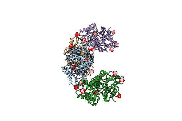
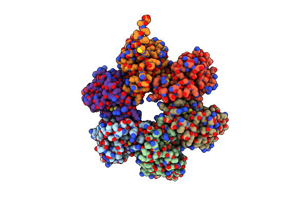
Crystal Structure Of Cystathionine Gamma-Lyase From Lactobacillus Plantarum Complexed With L-Serine
Organism: Lactobacillus plantarum
Method: X-RAY DIFFRACTION Resolution:2.75 Å Release Date: 2020-10-07 Classification: LYASE Ligands: KOU, PO4 |
 |
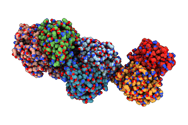
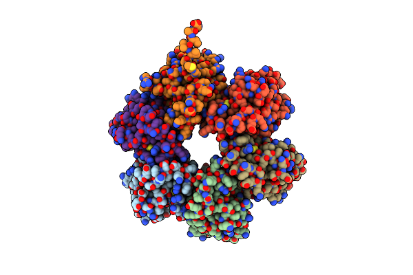
Crystal Structure Of Cystathionine Gamma-Lyase From Lactobacillus Plantarum Complexed With Cystathionine
Organism: Lactobacillus plantarum
Method: X-RAY DIFFRACTION Resolution:3.10 Å Release Date: 2020-10-07 Classification: LYASE Ligands: E9U, PO4 |
 |
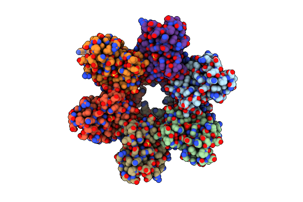
Crystal Structure Of Cystathionine Beta-Synthase From Lactobacillus Plantarum
Organism: Lactobacillus plantarum wcfs1
Method: X-RAY DIFFRACTION Resolution:2.40 Å Release Date: 2016-12-07 Classification: LYASE Ligands: SO4, GOL |
 |
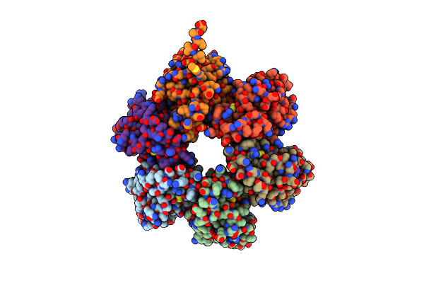
Crystal Structure Of K42A Mutant Of Cystathionine Beta-Synthase From Lactobacillus Plantarum In A Complex With L-Methionine
Organism: Lactobacillus plantarum wcfs1
Method: X-RAY DIFFRACTION Resolution:3.30 Å Release Date: 2016-12-07 Classification: LYASE Ligands: AA5, SO4 |
 |
Organism: Escherichia coli
Method: X-RAY DIFFRACTION Resolution:1.82 Å Release Date: 2014-11-12 Classification: HYDROLASE Ligands: SO4 |
 |
Organism: Escherichia coli
Method: X-RAY DIFFRACTION Resolution:1.70 Å Release Date: 2014-11-12 Classification: HYDROLASE Ligands: SO4 |
 |
Organism: Escherichia coli
Method: X-RAY DIFFRACTION Resolution:2.07 Å Release Date: 2014-11-12 Classification: HYDROLASE Ligands: SO4 |
 |
Organism: Escherichia coli
Method: X-RAY DIFFRACTION Resolution:1.80 Å Release Date: 2014-11-12 Classification: HYDROLASE Ligands: SO4 |