
Search Count: 785
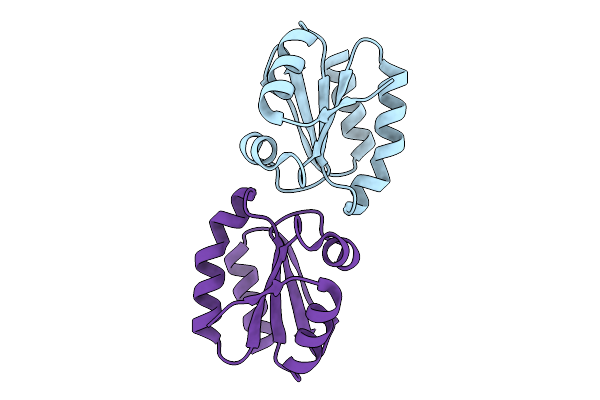 |
Organism: Rattus norvegicus
Method: X-RAY DIFFRACTION Release Date: 2025-09-24 Classification: ELECTRON TRANSPORT |
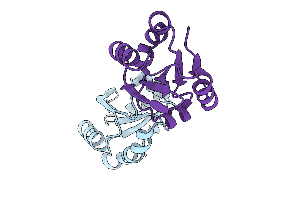 |
Organism: Rattus norvegicus
Method: X-RAY DIFFRACTION Release Date: 2025-09-24 Classification: ELECTRON TRANSPORT |
 |
Organism: Deinococcus radiodurans r1 = atcc 13939 = dsm 20539
Method: X-RAY DIFFRACTION Release Date: 2025-09-24 Classification: METAL BINDING PROTEIN Ligands: CU, GOL |
 |
Organism: Deinococcus radiodurans r1 = atcc 13939 = dsm 20539
Method: X-RAY DIFFRACTION Release Date: 2025-09-24 Classification: METAL BINDING PROTEIN Ligands: ZN, CU, CA, SO4, CL |
 |
Organism: Gluconobacter oxydans
Method: X-RAY DIFFRACTION Release Date: 2025-09-03 Classification: TRANSFERASE Ligands: EDO, CIT |
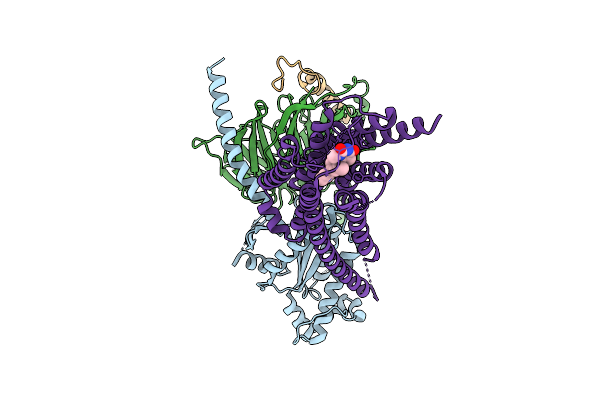 |
Organism: Homo sapiens
Method: ELECTRON MICROSCOPY Release Date: 2025-07-16 Classification: HORMONE Ligands: A1IZU |
 |
Organism: Homo sapiens
Method: ELECTRON MICROSCOPY Release Date: 2025-07-16 Classification: HORMONE Ligands: A1IZV |
 |
Organism: Bacillus sp. ps3
Method: ELECTRON MICROSCOPY Release Date: 2025-07-09 Classification: MEMBRANE PROTEIN Ligands: ADP, MG |
 |
Organism: Bacillus sp. ps3
Method: ELECTRON MICROSCOPY Release Date: 2025-07-09 Classification: MEMBRANE PROTEIN |
 |
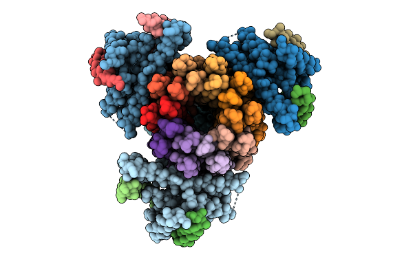
Organism: Homo sapiens
Method: ELECTRON MICROSCOPY Release Date: 2025-06-11 Classification: NUCLEAR PROTEIN Ligands: ATP, MG, ADP |
 |
Organism: Homo sapiens
Method: ELECTRON MICROSCOPY Release Date: 2025-06-11 Classification: NUCLEAR PROTEIN Ligands: ZN, ATP, MG, ADP |
 |
Organism: Homo sapiens
Method: ELECTRON MICROSCOPY Release Date: 2025-06-11 Classification: NUCLEAR PROTEIN Ligands: ZN, ATP, ADP |
 |
Organism: Homo sapiens
Method: ELECTRON MICROSCOPY Release Date: 2025-06-11 Classification: NUCLEAR PROTEIN Ligands: ATP, MG, ADP |
 |
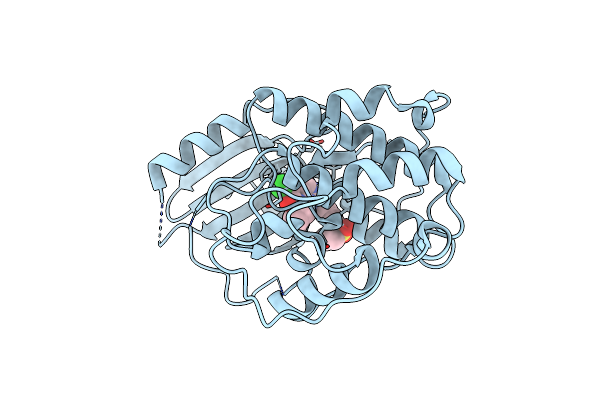
Crystal Structure Of The Enzyme-Product Complex Of L-Azetidine-2-Carboxylate Hydrolase
Organism: Pseudomonas sp. a2c
Method: X-RAY DIFFRACTION Resolution:0.93 Å Release Date: 2025-06-04 Classification: HYDROLASE Ligands: 42B, IMD, FMT, EDO, PEG, MG |
 |
Organism: Homo sapiens
Method: X-RAY DIFFRACTION Resolution:2.60 Å Release Date: 2025-04-16 Classification: CELL CYCLE Ligands: A1A1H |
 |
Organism: Homo sapiens
Method: X-RAY DIFFRACTION Resolution:2.54 Å Release Date: 2025-04-16 Classification: CELL CYCLE Ligands: A1A1H, PEG, GOL, EDO |
 |
Organism: Homo sapiens
Method: ELECTRON MICROSCOPY Release Date: 2025-04-16 Classification: CELL CYCLE Ligands: ZN, A1A1I |
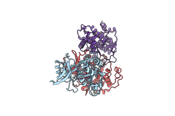 |
Cryo-Em Structure Of Cdk2/Cycline1 In Complex With Crbn/Ddb1 And Cpd 4 (Local Mask)
Organism: Homo sapiens
Method: ELECTRON MICROSCOPY Release Date: 2025-04-16 Classification: CELL CYCLE Ligands: ZN, A1A1I |
 |
Organism: Homo sapiens
Method: ELECTRON MICROSCOPY Release Date: 2025-02-26 Classification: MEMBRANE PROTEIN Ligands: GLY, NAG, GLU |
 |
Organism: Homo sapiens
Method: ELECTRON MICROSCOPY Release Date: 2025-02-26 Classification: MEMBRANE PROTEIN Ligands: GLY, NAG, GLU |