
Search Count: 14
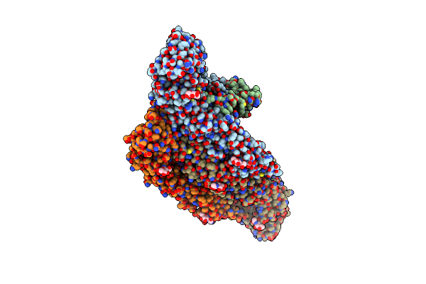 |
Organism: Usutu virus
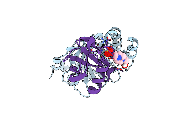
Method: ELECTRON MICROSCOPY Resolution:2.42 Å Release Date: 2021-09-01 Classification: VIRUS Ligands: 0SM, PC7, NAG |
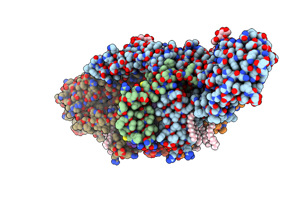 |
Organism: Usutu virus
Method: ELECTRON MICROSCOPY Resolution:2.35 Å Release Date: 2021-09-01 Classification: VIRUS Ligands: 0SM, PC7, NAG |
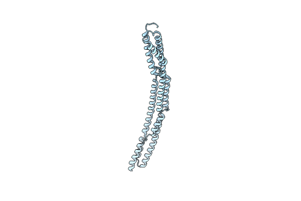 |
Novel Structure Of The N-Terminal Helical Domain Of Biba, A Group B Streptococcus Immunogenic Bacterial Adhesin
Organism: Streptococcus agalactiae
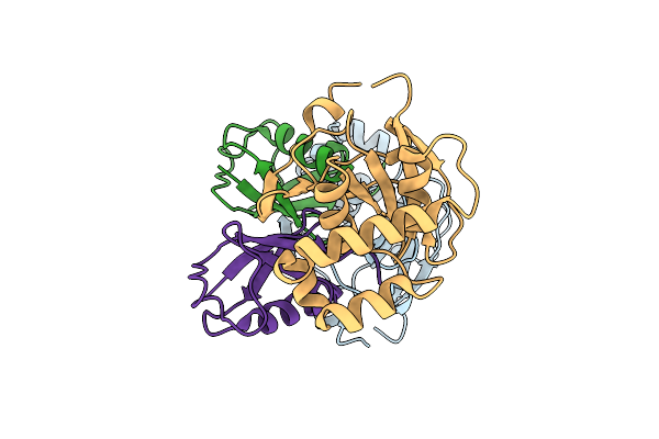
Method: X-RAY DIFFRACTION Resolution:3.03 Å Release Date: 2020-08-12 Classification: IMMUNE SYSTEM |
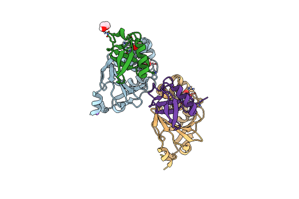 |
Otu Protease Of Crimean Congo Hemorrhagic Fever Virus Bound To Ubiquitin Variant Cc.4
Organism: Crimean-congo hemorrhagic fever virus (strain nigeria/ibar10200/1970), Synthetic construct
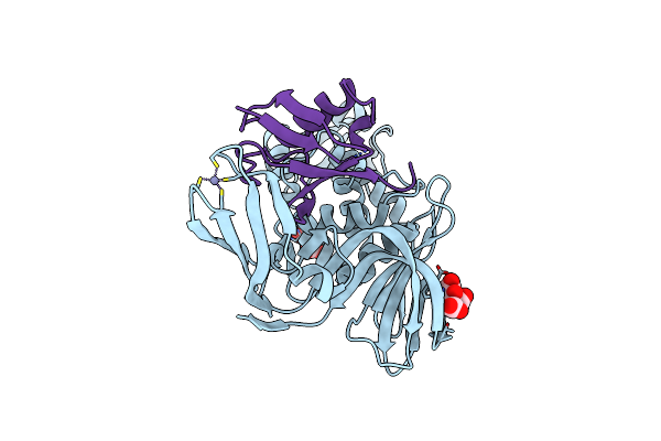
Method: X-RAY DIFFRACTION Resolution:2.10 Å Release Date: 2017-05-10 Classification: hydrolase, transferase Ligands: EDO, NA |
 |
Otu Protease Of Crimean Congo Hemorrhagic Fever Virus Bound To Ubiquitin Variant Cc.2
Organism: Crimean-congo hemorrhagic fever virus (strain nigeria/ibar10200/1970), Synthetic construct
Method: X-RAY DIFFRACTION Resolution:1.50 Å Release Date: 2017-05-10 Classification: hydrolase, transferase |
 |
Otu Protease Of Crimean Congo Hemorrhagic Fever Virus Bound To Ubiquitin Variant Cc.1
Organism: Crimean-congo hemorrhagic fever virus (strain nigeria/ibar10200/1970), Synthetic construct
Method: X-RAY DIFFRACTION Resolution:2.20 Å Release Date: 2017-05-10 Classification: hydrolase, transferase |
 |
Crystal Structure Of The Middle East Respiratory Syndrome Coronavirus Papain-Like Protease Bound To Ubiquitin Variant Me.4
Organism: Human betacoronavirus 2c emc/2012, Homo sapiens
Method: X-RAY DIFFRACTION Resolution:2.55 Å Release Date: 2017-05-10 Classification: HYDROLASE Ligands: ZN, CL, NA, PGO |
 |
Crystal Structure Of The Middle East Respiratory Syndrome Coronavirus Papain-Like Protease Bound To Ubiquitin Variant Me.2
Organism: Human betacoronavirus 2c emc/2012, Homo sapiens
Method: X-RAY DIFFRACTION Resolution:2.70 Å Release Date: 2017-05-10 Classification: HYDROLASE Ligands: ZN, FLC, CL |
 |
Organism: Streptococcus agalactiae serogroup v
Method: X-RAY DIFFRACTION Resolution:2.45 Å Release Date: 2011-10-26 Classification: HYDROLASE |
 |
The Crystal Structure Of The Complex Of Streptococcus Agalactiae Sortase C1 And Mtset
Organism: Streptococcus agalactiae serogroup v
Method: X-RAY DIFFRACTION Resolution:2.85 Å Release Date: 2011-10-26 Classification: HYDROLASE Ligands: ETM, SO4, CL |
 |
Organism: Streptococcus agalactiae serogroup v
Method: X-RAY DIFFRACTION Resolution:3.00 Å Release Date: 2011-09-07 Classification: HYDROLASE Ligands: SO4 |
 |
Organism: Streptococcus agalactiae serogroup v
Method: X-RAY DIFFRACTION Resolution:2.30 Å Release Date: 2011-09-07 Classification: HYDROLASE |
 |
Organism: Streptococcus agalactiae serogroup v
Method: X-RAY DIFFRACTION Resolution:2.90 Å Release Date: 2011-09-07 Classification: HYDROLASE Ligands: SO4 |
 |
Organism: Streptococcus agalactiae serogroup v
Method: X-RAY DIFFRACTION Resolution:3.10 Å Release Date: 2011-09-07 Classification: HYDROLASE Ligands: ZN |