
Search Count: 22
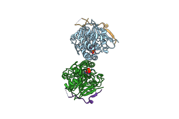 |
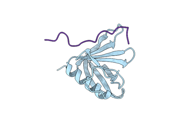
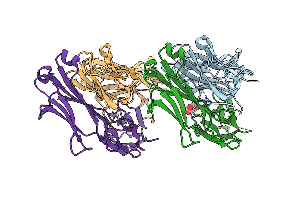
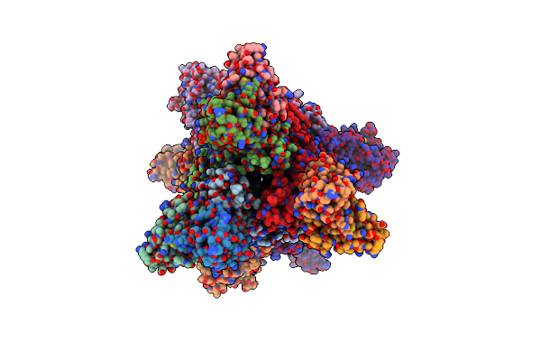
Protein Phosphatase 1 In Complex With Pp1-Specific Phosphatase Targeting Peptide (Phostap) Version 1
Organism: Homo sapiens
Method: X-RAY DIFFRACTION Resolution:2.39 Å Release Date: 2024-05-29 Classification: BIOSYNTHETIC PROTEIN Ligands: MN, PO4 |
 |
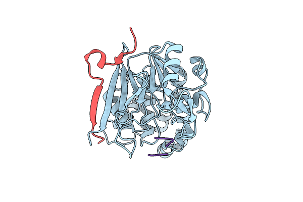
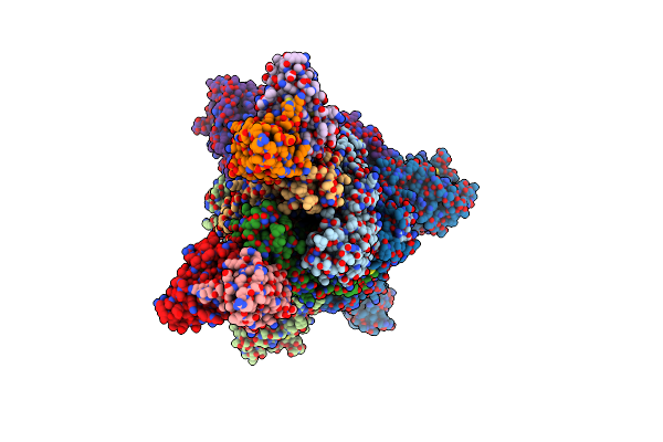
Protein Phosphatase 1 In Complex With Pp1-Specific Phosphatase Targeting Peptide (Phostap) Version 3
Organism: Homo sapiens
Method: X-RAY DIFFRACTION Resolution:1.76 Å Release Date: 2024-05-29 Classification: BIOSYNTHETIC PROTEIN Ligands: MN, SO4, CL |
 |
Organism: Homo sapiens
Method: X-RAY DIFFRACTION Resolution:1.59 Å Release Date: 2024-05-08 Classification: BIOSYNTHETIC PROTEIN Ligands: SO4, GOL |
 |
Organism: Homo sapiens
Method: X-RAY DIFFRACTION Resolution:3.15 Å Release Date: 2020-03-25 Classification: HYDROLASE |
 |
Organism: Homo sapiens
Method: X-RAY DIFFRACTION Resolution:2.45 Å Release Date: 2020-03-25 Classification: HYDROLASE |
 |
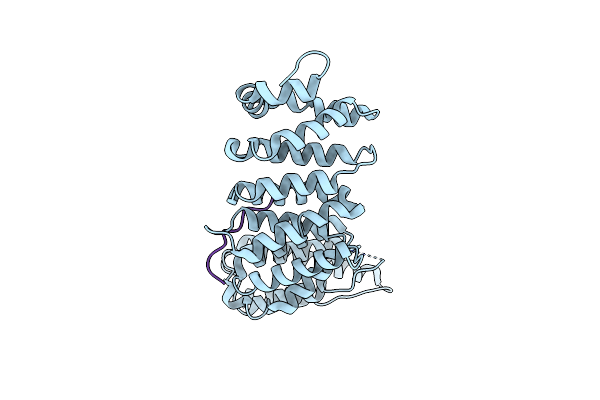
Organism: Homo sapiens
Method: X-RAY DIFFRACTION Resolution:1.52 Å Release Date: 2019-10-16 Classification: PEPTIDE BINDING PROTEIN |
 |
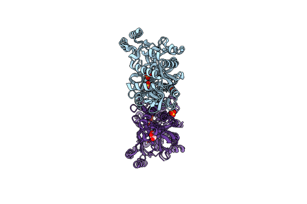
Crystal Structure Of Protein Phosphatase 1 (Pp1) Bound To The Muscle Glycogen-Targeting Subunit (Gm)
Organism: Homo sapiens, Oryctolagus cuniculus, Synthetic construct
Method: X-RAY DIFFRACTION Resolution:1.45 Å Release Date: 2019-01-23 Classification: SIGNALING PROTEIN |
 |
Crystal Structure Of Protein Phosphatase 1 Bound To The Natural Inhibitor Tautomycetin
Organism: Homo sapiens
Method: X-RAY DIFFRACTION Resolution:2.21 Å Release Date: 2017-11-29 Classification: HYDROLASE/HYDROLASE INHIBITOR Ligands: MN, BKM, DMS, CL |
 |
Organism: Middle east respiratory syndrome-related coronavirus
Method: X-RAY DIFFRACTION Resolution:2.00 Å Release Date: 2017-08-30 Classification: VIRAL PROTEIN Ligands: NAG, FOL, MPD, IMD |
 |
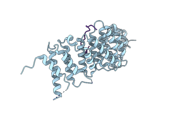
Organism: Mus musculus
Method: X-RAY DIFFRACTION Resolution:1.57 Å Release Date: 2017-08-30 Classification: IMMUNE SYSTEM Ligands: GOL |
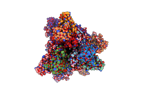 |
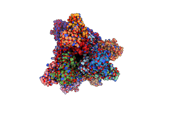
Mers S Ectodomain Trimer In Complex With Variable Domain Of Neutralizing Antibody G4
Organism: Middle east respiratory syndrome-related coronavirus, Mus musculus
Method: ELECTRON MICROSCOPY Release Date: 2017-08-16 Classification: VIRAL PROTEIN Ligands: NAG |
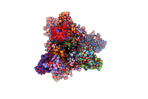 |
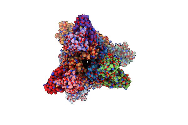
Mers S Ectodomain Trimer In Complex With Variable Domain Of Neutralizing Antibody G4
Organism: Middle east respiratory syndrome-related coronavirus, Mus musculus
Method: ELECTRON MICROSCOPY Release Date: 2017-08-16 Classification: VIRAL PROTEIN Ligands: NAG |
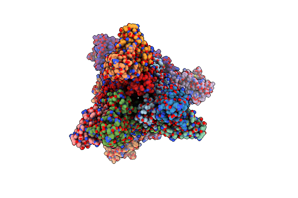 |
Mers S Ectodomain Trimer In Complex With Variable Domain Of Neutralizing Antibody G4
Organism: Middle east respiratory syndrome-related coronavirus, Mus musculus
Method: ELECTRON MICROSCOPY Release Date: 2017-08-16 Classification: VIRAL PROTEIN |
 |
Mers S Ectodomain Trimer In Complex With Variable Domain Of Neutralizing Antibody G4
Organism: Middle east respiratory syndrome-related coronavirus, Mus musculus
Method: ELECTRON MICROSCOPY Release Date: 2017-08-16 Classification: VIRAL PROTEIN |
 |
Mers S Ectodomain Trimer In Complex With Variable Domain Of Neutralizing Antibody G4
Organism: Middle east respiratory syndrome-related coronavirus, Mus musculus
Method: ELECTRON MICROSCOPY Release Date: 2017-08-16 Classification: VIRAL PROTEIN |
 |
Mers S Ectodomain Trimer In Complex With Variable Domain Of Neutralizing Antibody G4
Organism: Middle east respiratory syndrome-related coronavirus, Mus musculus
Method: ELECTRON MICROSCOPY Release Date: 2017-08-16 Classification: VIRAL PROTEIN |
 |
Mers S Ectodomain Trimer In Complex With Variable Domain Of Neutralizing Antibody G4
Organism: Middle east respiratory syndrome-related coronavirus, Mus musculus
Method: ELECTRON MICROSCOPY Release Date: 2017-08-16 Classification: VIRAL PROTEIN |
 |
Mers S Ectodomain Trimer In Complex With Variable Domain Of Neutralizing Antibody G4
Organism: Middle east respiratory syndrome-related coronavirus, Mus musculus
Method: ELECTRON MICROSCOPY Release Date: 2017-08-16 Classification: VIRAL PROTEIN |
 |
Mers S Ectodomain Trimer In Complex With Variable Domain Of Neutralizing Antibody G4
Organism: Middle east respiratory syndrome-related coronavirus, Mus musculus
Method: ELECTRON MICROSCOPY Release Date: 2017-08-16 Classification: VIRAL PROTEIN |
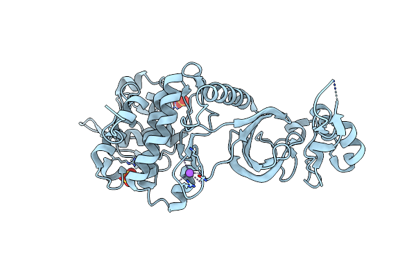 |
Crystal Structure Of Dual-Specificity Tyrosine Phosphorylation Regulated Kinase 2 (Dyrk2)
Organism: Homo sapiens
Method: X-RAY DIFFRACTION Resolution:2.36 Å Release Date: 2009-10-13 Classification: TRANSFERASE Ligands: SO4, NA, CL |