
Search Count: 16
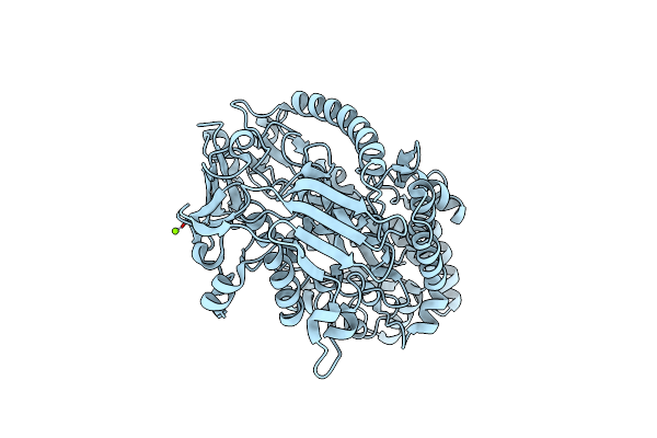 |
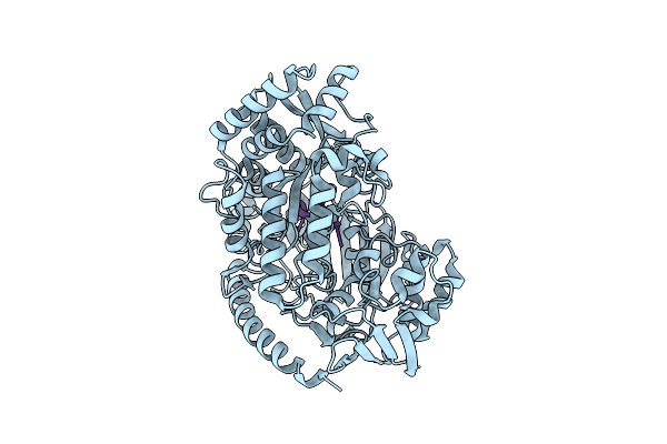
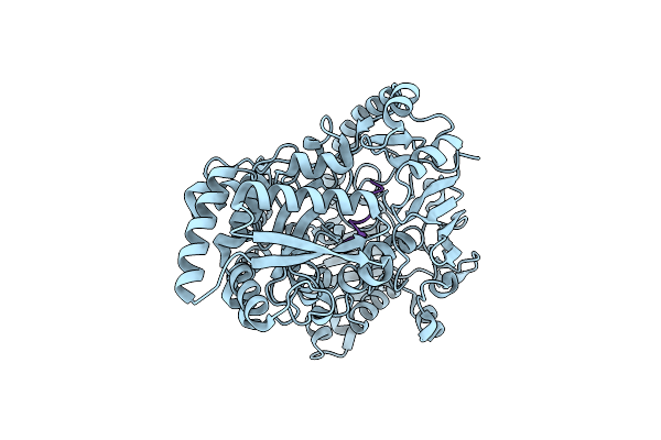
Crystal Structure Of The Pneumococcal Substrate-Binding Protein Alid In Open Conformation
Organism: Streptococcus pneumoniae
Method: X-RAY DIFFRACTION Resolution:1.80 Å Release Date: 2024-05-22 Classification: PEPTIDE BINDING PROTEIN Ligands: MG |
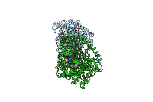 |
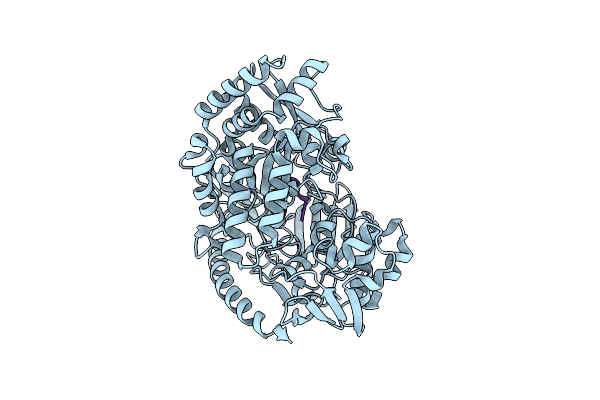
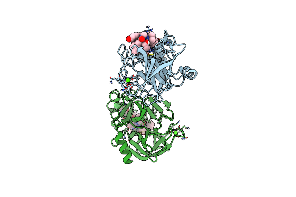
Crystal Structure Of The Pneumococcal Substrate-Binding Protein Alid In Closed Conformation In Complex With Peptide 1
Organism: Streptococcus pneumoniae, Prevotella
Method: X-RAY DIFFRACTION Resolution:2.10 Å Release Date: 2024-05-22 Classification: PEPTIDE BINDING PROTEIN Ligands: ZN |
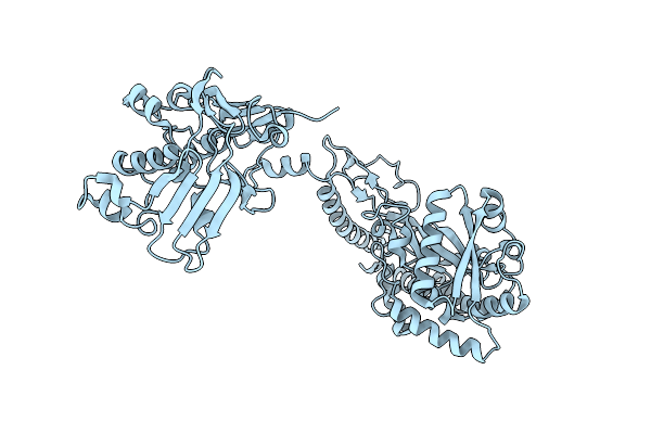 |
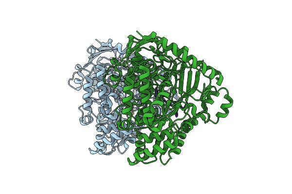
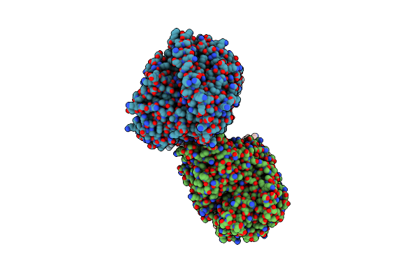
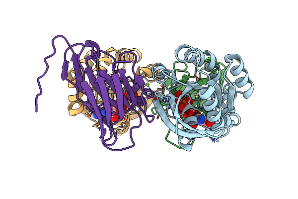
Crystal Structure Of The Pneumococcal Substrate-Binding Protein Alic As A Domain-Swapped Dimer
Organism: Streptococcus pneumoniae
Method: X-RAY DIFFRACTION Resolution:2.38 Å Release Date: 2024-05-22 Classification: PEPTIDE BINDING PROTEIN |
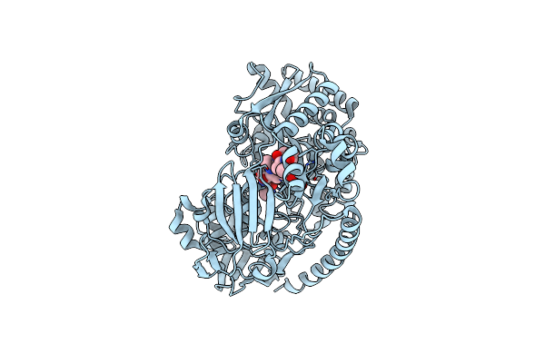 |
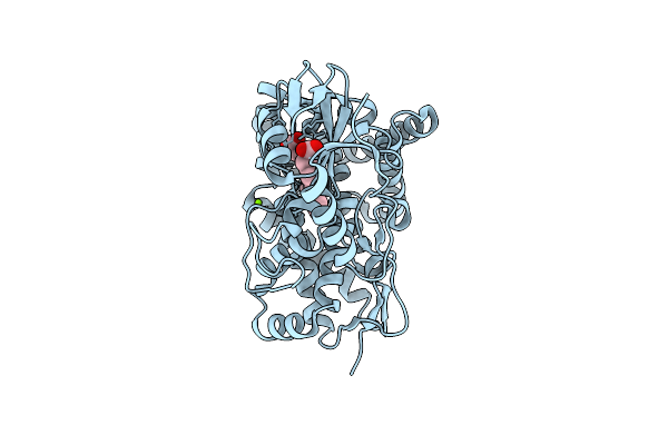
Crystal Structure Of The Pneumococcal Substrate-Binding Protein Alib In Complex With An Unknown Peptide
Organism: Streptococcus pneumoniae, Escherichia coli bl21(de3)
Method: X-RAY DIFFRACTION Resolution:1.65 Å Release Date: 2024-05-22 Classification: PEPTIDE BINDING PROTEIN |
 |
Crystal Structure Of The Pneumococcal Substrate-Binding Protein Alib In Complex With Peptide 2
Organism: Streptococcus pneumoniae, Synthetic construct
Method: X-RAY DIFFRACTION Resolution:2.29 Å Release Date: 2024-05-22 Classification: PEPTIDE BINDING PROTEIN |
 |
Crystal Structure Of The Pneumococcal Substrate-Binding Protein Alib In Complex With Peptide 3
Organism: Streptococcus pneumoniae, Synthetic construct
Method: X-RAY DIFFRACTION Resolution:1.66 Å Release Date: 2024-05-22 Classification: PEPTIDE BINDING PROTEIN |
 |
Crystal Structure Of The Pneumococcal Substrate-Binding Protein Alib In Complex With Peptide 4
Organism: Streptococcus pneumoniae, Synthetic construct
Method: X-RAY DIFFRACTION Resolution:1.49 Å Release Date: 2024-05-22 Classification: PEPTIDE BINDING PROTEIN |
 |
Crystal Structure Of The Pneumococcal Substrate-Binding Protein Amia In Complex With Peptide 5
Organism: Streptococcus pneumoniae, Bacteria
Method: X-RAY DIFFRACTION Resolution:1.76 Å Release Date: 2024-05-22 Classification: PEPTIDE BINDING PROTEIN |
 |
Crystal Structure Of The Pneumococcal Substrate-Binding Protein Amia In Complex With An Unknown Peptide
Organism: Streptococcus pneumoniae, Escherichia coli
Method: X-RAY DIFFRACTION Resolution:1.50 Å Release Date: 2023-12-20 Classification: PEPTIDE BINDING PROTEIN |
 |
Crystal Structure Of Fructose-Bisphosphate Aldolases Fbac From Bacillus Methanolicus
Organism: Bacillus methanolicus (strain mga3 / atcc 53907)
Method: X-RAY DIFFRACTION Resolution:2.20 Å Release Date: 2021-02-17 Classification: LYASE Ligands: 13P |
 |
Crystal Structure Of Fructose-Bisphosphate Aldolase Fbap From Bacillus Methanolicus
Organism: Bacillus methanolicus (strain mga3 / atcc 53907)
Method: X-RAY DIFFRACTION Resolution:2.00 Å Release Date: 2021-02-17 Classification: LYASE Ligands: MLT, PO4, IMD |
 |
Organism: Synthetic construct, Sus scrofa
Method: X-RAY DIFFRACTION Resolution:2.20 Å Release Date: 2020-03-04 Classification: HYDROLASE Ligands: CA |
 |
Organism: Homo sapiens
Method: X-RAY DIFFRACTION Resolution:1.19 Å Release Date: 2019-09-25 Classification: IMMUNE SYSTEM Ligands: GTP, MG |
 |
Organism: Homo sapiens
Method: X-RAY DIFFRACTION Resolution:1.50 Å Release Date: 2019-09-25 Classification: IMMUNE SYSTEM Ligands: GTP, MG |
 |
Organism: Sorangium cellulosum (strain so ce56)
Method: X-RAY DIFFRACTION Resolution:1.85 Å Release Date: 2016-08-03 Classification: OXIDOREDUCTASE Ligands: HEM, MG |
 |
Organism: Saccharomyces cerevisiae
Method: X-RAY DIFFRACTION Resolution:2.80 Å Release Date: 2015-02-18 Classification: HYDROLASE Ligands: MG, CL, 7IM, MES |