
Search Count: 78
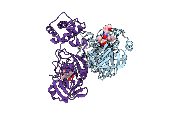 |
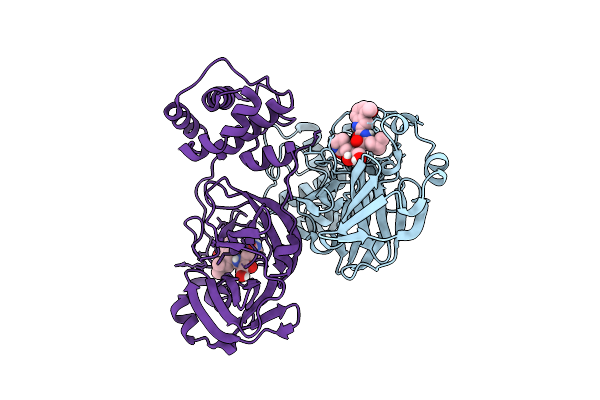
Crystal Structure Of Sars-Cov-2 Mpro S10A In Complex With Pfizer Intravenous Inhibitor Pf-00835231
Organism: Severe acute respiratory syndrome coronavirus 2
Method: X-RAY DIFFRACTION Release Date: 2025-12-10 Classification: VIRAL PROTEIN Ligands: V2M |
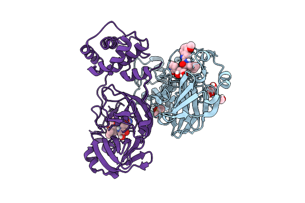 |
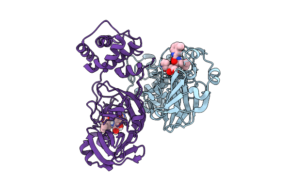
Crystal Structure Of Sars-Cov-2 Mpro S10C In Complex With Pfizer Intravenous Inhibitor Pf-00835231
Organism: Severe acute respiratory syndrome coronavirus 2
Method: X-RAY DIFFRACTION Release Date: 2025-12-10 Classification: VIRAL PROTEIN Ligands: DMS, EDO, V2M |
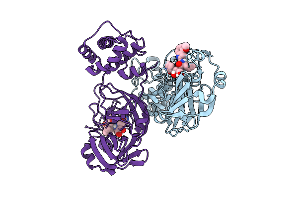 |
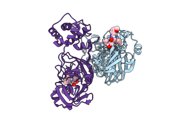
Crystal Structure Of Sars-Cov-2 Mpro S113A In Complex With Pfizer Intravenous Inhibitor Pf-00835231
Organism: Severe acute respiratory syndrome coronavirus 2
Method: X-RAY DIFFRACTION Release Date: 2025-12-10 Classification: VIRAL PROTEIN Ligands: V2M |
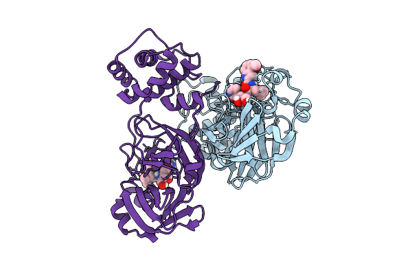 |
Crystal Structure Of Sars-Cov-2 Mpro S113C In Complex With Pfizer Intravenous Inhibitor Pf-00835231
Organism: Severe acute respiratory syndrome coronavirus 2
Method: X-RAY DIFFRACTION Release Date: 2025-12-10 Classification: VIRAL PROTEIN Ligands: V2M |
 |
Crystal Structure Of Sars-Cov-2 Mpro L115A In Complex With Pfizer Intravenous Inhibitor Pf-00835231
Organism: Severe acute respiratory syndrome coronavirus 2
Method: X-RAY DIFFRACTION Release Date: 2025-12-10 Classification: VIRAL PROTEIN Ligands: V2M |
 |
Crystal Structure Of Sars-Cov-2 Mpro L115M In Complex With Pfizer Intravenous Inhibitor Pf-00835231
Organism: Severe acute respiratory syndrome coronavirus 2
Method: X-RAY DIFFRACTION Release Date: 2025-12-10 Classification: VIRAL PROTEIN Ligands: V2M |
 |
Crystal Structure Of Sars-Cov-2 Mpro S147A In Complex With Pfizer Intravenous Inhibitor Pf-00835231
Organism: Severe acute respiratory syndrome coronavirus 2
Method: X-RAY DIFFRACTION Release Date: 2025-12-10 Classification: VIRAL PROTEIN Ligands: V2M |
 |
Crystal Structure Of Sars-Cov-2 Mpro S147N In Complex With Pfizer Intravenous Inhibitor Pf-00835231
Organism: Severe acute respiratory syndrome coronavirus 2
Method: X-RAY DIFFRACTION Release Date: 2025-12-10 Classification: VIRAL PROTEIN Ligands: V2M |
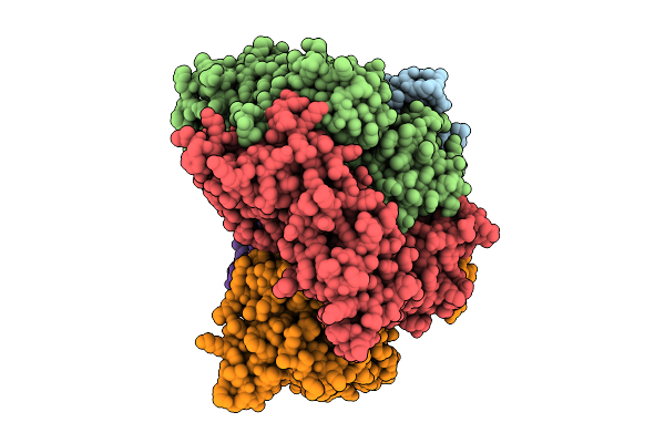 |
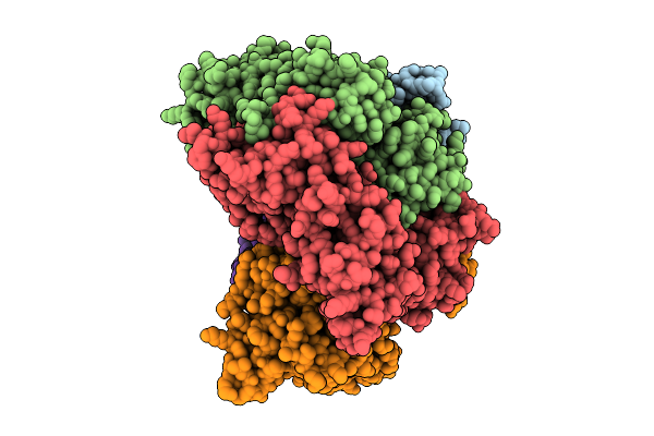
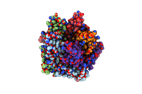
Structure Of The S. Cerevisiae Clamp Loader Replication Factor C (Rfc) With Mixed Nucleotide Occupancy
Organism: Saccharomyces cerevisiae
Method: ELECTRON MICROSCOPY Release Date: 2025-11-12 Classification: REPLICATION Ligands: ACT, ADP, GDP |
 |
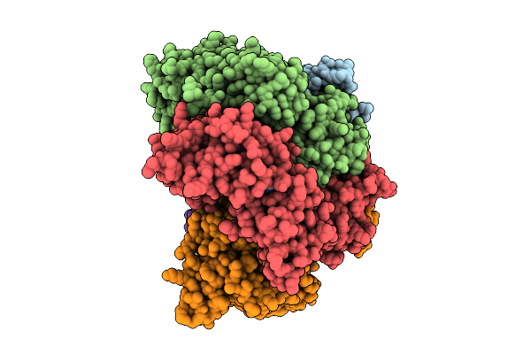
Structure Of The S. Cerevisiae Clamp Loader Replication Factor C (Rfc) With Mixed Nucleotide Occupancy
Organism: Saccharomyces cerevisiae
Method: ELECTRON MICROSCOPY Release Date: 2025-11-12 Classification: REPLICATION Ligands: ACT, ADP, GDP |
 |
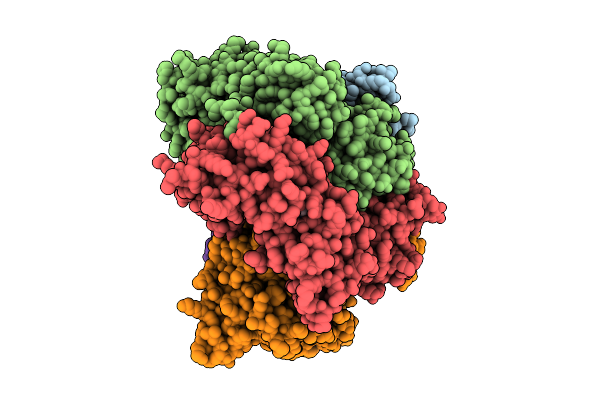
Structure Of The S. Cerevisiae Clamp Loader Replication Factor C (Rfc) With Mixed Nucleotide Occupancy
Organism: Saccharomyces cerevisiae
Method: ELECTRON MICROSCOPY Release Date: 2025-11-12 Classification: REPLICATION Ligands: ACT, ADP, GDP |
 |
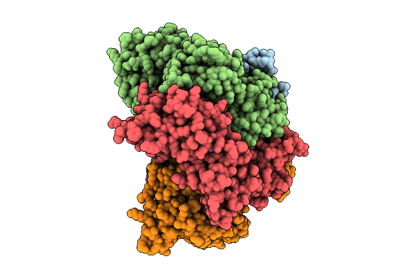
Structure Of The S. Cerevisiae Clamp Loader Replication Factor C (Rfc) With Mixed Nucleotide Occupancy
Organism: Saccharomyces cerevisiae
Method: ELECTRON MICROSCOPY Release Date: 2025-11-12 Classification: REPLICATION Ligands: ACT, ADP, GDP |
 |
Structure Of The S. Cerevisiae Clamp Loader Replication Factor C (Rfc) With Mixed Nucleotide Occupancy
Organism: Saccharomyces cerevisiae
Method: ELECTRON MICROSCOPY Release Date: 2025-11-12 Classification: REPLICATION Ligands: ACT, ADP, GDP |
 |
Structure Of The S. Cerevisiae Clamp Loader Replication Factor C (Rfc) With Mixed Nucleotide Occupancy
Organism: Saccharomyces cerevisiae
Method: ELECTRON MICROSCOPY Release Date: 2025-11-12 Classification: REPLICATION Ligands: ACT, ADP, GDP |
 |
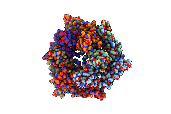
Structure Of The Five-Fold Capsomer Of The Drosophila Retrotransposon Copia Capsid
Organism: Drosophila
Method: ELECTRON MICROSCOPY Release Date: 2025-02-12 Classification: VIRUS LIKE PARTICLE |
 |
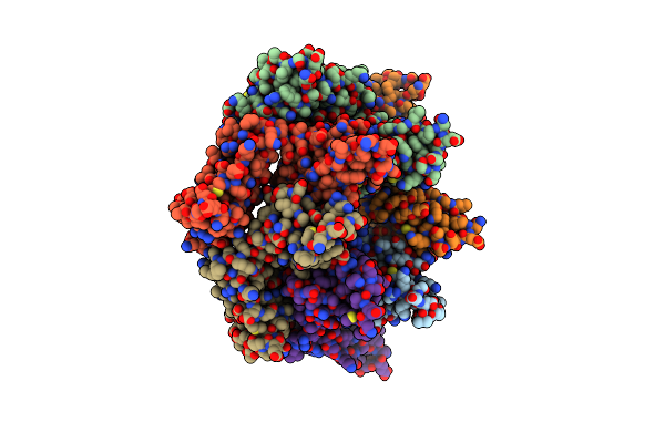
Structure Of The Three-Fold Capsomer Of The Drosophila Retrotransposon Copia Capsid
Organism: Drosophila
Method: ELECTRON MICROSCOPY Release Date: 2025-02-12 Classification: VIRUS LIKE PARTICLE |
 |
Structure Of The Non-Symmetric Capsomer Of The Drosophila Retrotransposon Copia Capsid
Organism: Drosophila
Method: ELECTRON MICROSCOPY Release Date: 2025-02-12 Classification: VIRUS LIKE PARTICLE |
 |
Organism: Drosophila
Method: ELECTRON MICROSCOPY Release Date: 2025-02-12 Classification: VIRUS LIKE PARTICLE |
 |
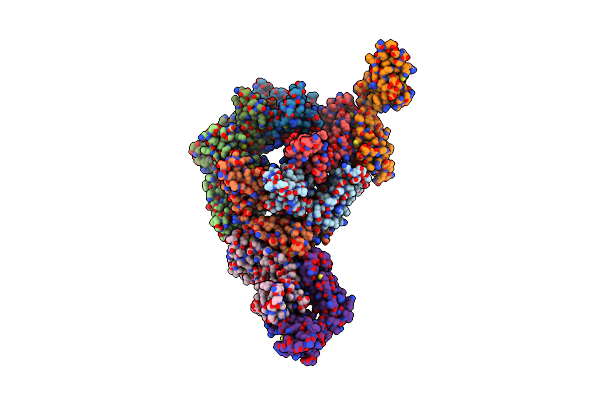
Structure Of The E. Coli Clamp Loader Bound To The Beta Clamp In A Open-Dnap/T Conformation
Organism: Escherichia coli, Synthetic construct
Method: ELECTRON MICROSCOPY Release Date: 2024-03-27 Classification: REPLICATION, TRANSFERASE/DNA Ligands: ZN, ADP, BEF, MG |
 |
Structure Of The E. Coli Clamp Loader Bound To The Beta Clamp In A Semi-Open Conformation
Organism: Escherichia coli
Method: ELECTRON MICROSCOPY Release Date: 2024-03-27 Classification: REPLICATION Ligands: ZN, ADP, BEF, MG |