
Search Count: 433
 |
Crystal Structure Of Iron-Bound Human Ado C18S/C239S Variant Soaked In Hydralazine At 2.39 Angstrom Resolution
Organism: Homo sapiens
Method: X-RAY DIFFRACTION Release Date: 2025-09-24 Classification: OXIDOREDUCTASE Ligands: FE2, HLZ, GOL |
 |
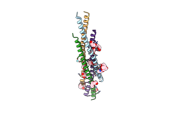
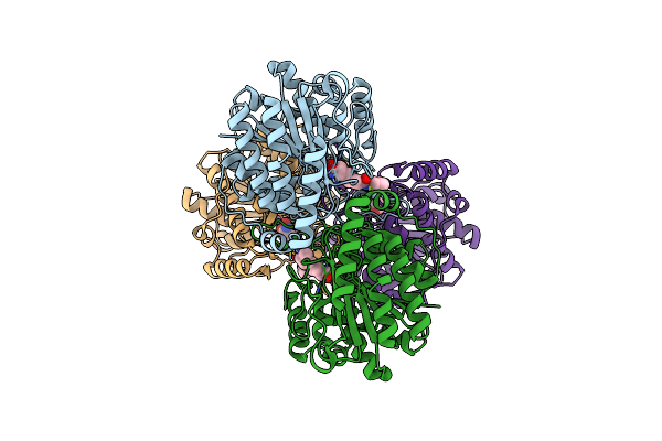
Crystal Structure Of Cobalt-Bound Human Ado C18S/C239S Variant In Complex With Hydralazine At 1.89 Angstrom Resolution
Organism: Homo sapiens
Method: X-RAY DIFFRACTION Release Date: 2025-09-10 Classification: OXIDOREDUCTASE Ligands: CO, HLZ, GOL, SO4 |
 |
Crystal Structure Of The Keap1 Kelch Domain In Complex With The Xchem Fragment Z19735904 At 1.14 Angstrom Resolution.
Organism: Mus musculus
Method: X-RAY DIFFRACTION Release Date: 2025-09-03 Classification: PEPTIDE BINDING PROTEIN Ligands: B0A, SO4, DMS |
 |
Crystal Structure Of The Keap1 Kelch Domain In Complex With The Small Molecule Ucab#827 At 1.40 Angstrom Resolution
Organism: Mus musculus
Method: X-RAY DIFFRACTION Release Date: 2025-09-03 Classification: PEPTIDE BINDING PROTEIN Ligands: A1IX2, CL, SO4, DMS |
 |
Crystal Structure Of The Keap1 Kelch Domain In Complex With The Small Molecule Ucab#909 At 1.61 Angstrom Resolution
Organism: Mus musculus
Method: X-RAY DIFFRACTION Release Date: 2025-09-03 Classification: PEPTIDE BINDING PROTEIN Ligands: A1IX3, SO4, DMS, CL |
 |
Crystal Structure Of The Keap1 Kelch Domain In Complex With The Small Molecule Ucab#985 At 1.65 Angstrom Resolution
Organism: Mus musculus
Method: X-RAY DIFFRACTION Release Date: 2025-09-03 Classification: PEPTIDE BINDING PROTEIN Ligands: A1IX4, SO4, DMS, CL |
 |
Crystal Structure Of The Keap1 Kelch Domain In Complex With The Small Molecule Ucab#1004 At 1.40 Angstrom Resolution
Organism: Mus musculus
Method: X-RAY DIFFRACTION Release Date: 2025-09-03 Classification: PEPTIDE BINDING PROTEIN Ligands: A1IXY, SO4, CL, DMS |
 |
Crystal Structure Of The Keap1 Kelch Domain In Complex With The Small Molecule Ucab#1010 At 1.50 Angstrom Resolution
Organism: Mus musculus
Method: X-RAY DIFFRACTION Release Date: 2025-09-03 Classification: PEPTIDE BINDING PROTEIN Ligands: A1IXZ, SO4, CL, DMS |
 |
Crystal Structure Of The Keap1 Kelch Domain In Complex With The Small Molecule Ucab#1032 At 1.61 Angstrom Resolution
Organism: Mus musculus
Method: X-RAY DIFFRACTION Release Date: 2025-09-03 Classification: PEPTIDE BINDING PROTEIN Ligands: A1IX0, SO4, DMS |
 |
Crystal Structure Of The Keap1 Kelch Domain In Complex With The Small Molecule Ucab#1090 At 1.74 Angstrom Resolution
Organism: Mus musculus
Method: X-RAY DIFFRACTION Release Date: 2025-09-03 Classification: PEPTIDE BINDING PROTEIN Ligands: A1IX1, SO4, DMS |
 |
Organism: Homo sapiens
Method: X-RAY DIFFRACTION Release Date: 2025-07-09 Classification: TRANSFERASE Ligands: A1IUI |
 |
Organism: Homo sapiens
Method: ELECTRON MICROSCOPY Release Date: 2025-02-12 Classification: TRANSFERASE Ligands: MN, UGA |
 |
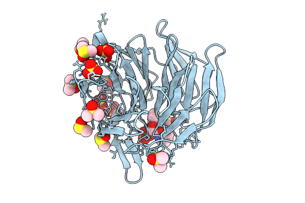
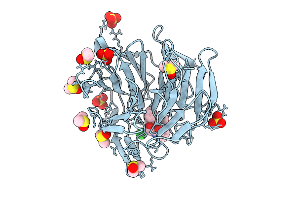
Organism: Homo sapiens
Method: X-RAY DIFFRACTION Resolution:1.44 Å Release Date: 2025-01-22 Classification: IMMUNE SYSTEM Ligands: TB, 9JE, BTB, PRO, YT3, PG4, ER3 |
 |
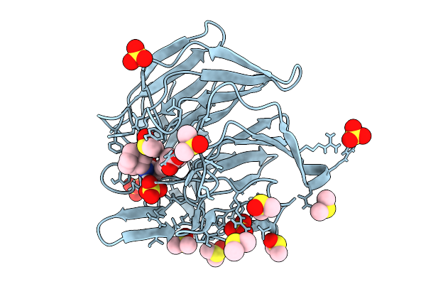
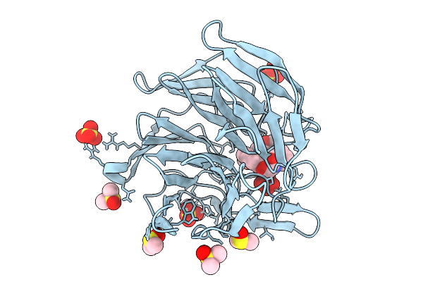
Tead1 In Complex With A Reversible Inhibitor N-[(1S)-2-Hydroxy-1-(1-Methyl-1H-Pyrazol-3-Yl)Ethyl]-2-Methyl-8-[4-(Trifluoromethyl)Phenyl]-2H,8H-Pyrazolo[3,4-B]Indole-5-Carboxamide
Organism: Homo sapiens
Method: X-RAY DIFFRACTION Resolution:1.93 Å Release Date: 2025-01-22 Classification: TRANSCRIPTION Ligands: GOL, A1IJL, SO4 |
 |
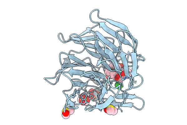
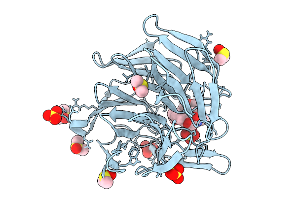
Organism: Plasmodium falciparum
Method: X-RAY DIFFRACTION Resolution:2.08 Å Release Date: 2024-11-06 Classification: SIGNALING PROTEIN Ligands: A1H19, GOL, EDO, SO4 |
 |
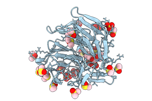
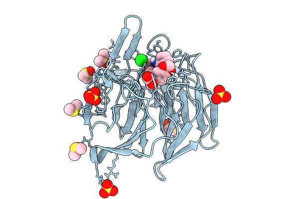
Organism: Homo sapiens
Method: X-RAY DIFFRACTION Release Date: 2024-11-06 Classification: IMMUNE SYSTEM Ligands: SO4 |
 |
Organism: Sphingomonas sp. y57
Method: X-RAY DIFFRACTION Resolution:1.48 Å Release Date: 2024-11-06 Classification: HYDROLASE Ligands: ACT, BGC, ZN, K |
 |
Crystal Structure Of Deacetylase (Hdah) From Vibrio Cholerae In Complex With Saha
Organism: Vibrio cholerae
Method: X-RAY DIFFRACTION Resolution:1.90 Å Release Date: 2024-11-06 Classification: HYDROLASE Ligands: SHH, ZN, K, ACT |
 |
Crystal Structure Of Dimethoate Hydrolase (Dmha) Of Rhizorhabdus Wittichii In Complex With Octanoic Acid
Organism: Rhizorhabdus wittichii dc-6
Method: X-RAY DIFFRACTION Resolution:2.10 Å Release Date: 2024-11-06 Classification: HYDROLASE Ligands: OCA, K, ZN, 1PE |
 |
Crystal Structure Of Rhizorhabdus Wittichii Dimethoate Hydrolase (Dmha) In Complex With Saha
Organism: Rhizorhabdus wittichii dc-6
Method: X-RAY DIFFRACTION Resolution:1.75 Å Release Date: 2024-11-06 Classification: HYDROLASE Ligands: SHH, ZN, K, OCA |