Search Count: 15
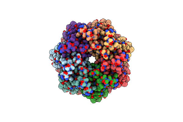 |
Organism: Xenopus tropicalis
Method: ELECTRON MICROSCOPY Release Date: 2024-12-11 Classification: MEMBRANE PROTEIN |
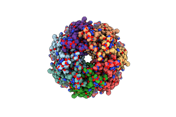 |
Organism: Xenopus tropicalis
Method: ELECTRON MICROSCOPY Release Date: 2024-12-04 Classification: MEMBRANE PROTEIN |
 |
Organism: Xenopus tropicalis
Method: ELECTRON MICROSCOPY Release Date: 2020-02-26 Classification: TRANSPORT PROTEIN |
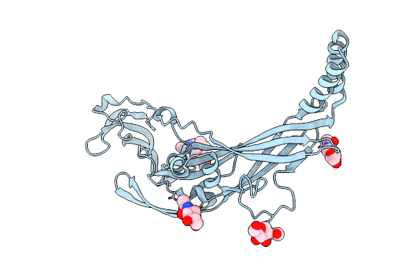 |
Crystal Structure Of The Atp-Gated P2X7 Ion Channel Bound To Allosteric Antagonist A740003
Organism: Ailuropoda melanoleuca
Method: X-RAY DIFFRACTION Resolution:3.60 Å Release Date: 2017-01-11 Classification: MEMBRANE PROTEIN Ligands: NAG, 7S4 |
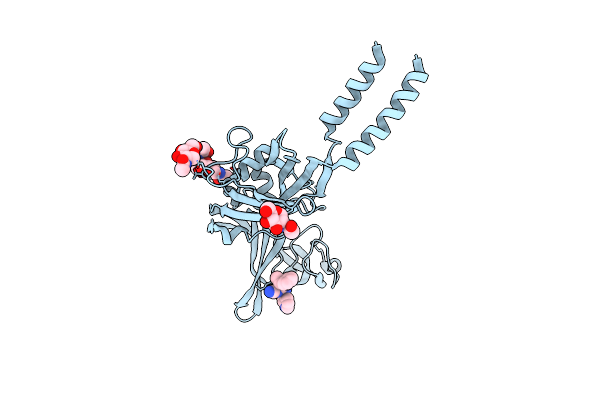 |
Crystal Structure Of The Atp-Gated P2X7 Ion Channel Bound To Allosteric Antagonist A804598
Organism: Ailuropoda melanoleuca
Method: X-RAY DIFFRACTION Resolution:3.40 Å Release Date: 2017-01-11 Classification: MEMBRANE PROTEIN Ligands: NAG, 7S1 |
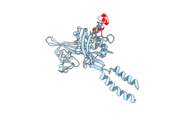 |
Crystal Structure Of The Atp-Gated P2X7 Ion Channel In The Closed, Apo State
Organism: Ailuropoda melanoleuca
Method: X-RAY DIFFRACTION Resolution:3.40 Å Release Date: 2017-01-04 Classification: MEMBRANE PROTEIN Ligands: NAG |
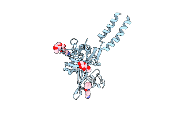 |
Crystal Structure Of The Atp-Gated P2X7 Ion Channel Bound To Allosteric Antagonist Az10606120
Organism: Ailuropoda melanoleuca
Method: X-RAY DIFFRACTION Resolution:3.50 Å Release Date: 2017-01-04 Classification: MEMBRANE PROTEIN Ligands: NAG, 7RY |
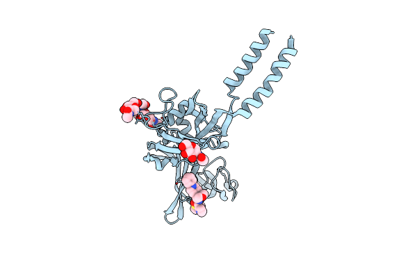 |
Crystal Structure Of The Atp-Gated P2X7 Ion Channel Bound To Allosteric Antagonist Jnj47965567
Organism: Ailuropoda melanoleuca
Method: X-RAY DIFFRACTION Resolution:3.20 Å Release Date: 2017-01-04 Classification: MEMBRANE PROTEIN Ligands: NAG, 7RV |
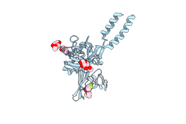 |
Crystal Structure Of The Atp-Gated P2X7 Ion Channel Bound To Allosteric Antagonist Gw791343
Organism: Ailuropoda melanoleuca
Method: X-RAY DIFFRACTION Resolution:3.30 Å Release Date: 2017-01-04 Classification: MEMBRANE PROTEIN Ligands: NAG, 7RS |
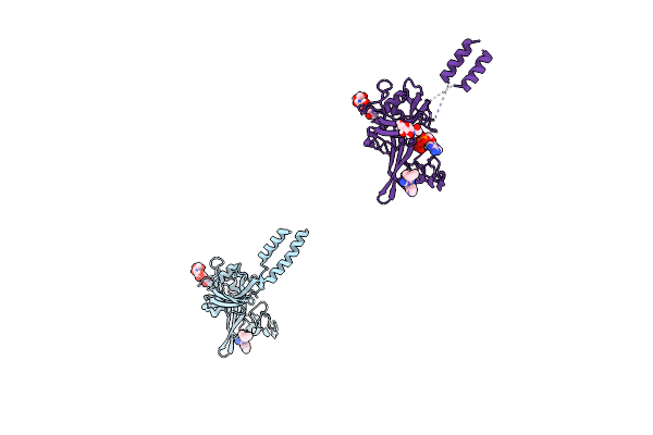 |
Crystal Structure Of The Atp-Gated P2X7 Ion Channel Bound To Atp And Allosteric Antagonist A804598
Organism: Ailuropoda melanoleuca
Method: X-RAY DIFFRACTION Resolution:3.90 Å Release Date: 2017-01-04 Classification: MEMBRANE PROTEIN Ligands: 7S1, NAG, ATP |
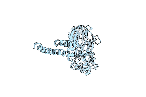 |
Organism: Gallus gallus
Method: X-RAY DIFFRACTION Resolution:3.00 Å Release Date: 2014-02-12 Classification: TRANSPORT PROTEIN Ligands: CL |
 |
Crystal Structure Of The Atp-Gated P2X4 Ion Channel In The Closed, Apo State At 3.5 Angstroms (R3)
Organism: Danio rerio
Method: X-RAY DIFFRACTION Resolution:3.46 Å Release Date: 2009-08-04 Classification: TRANSPORT PROTEIN Ligands: NAG |
 |
Crystal Structure Of The Atp-Gated P2X4 Ion Channel In The Closed, Apo State At 3.1 Angstroms
Organism: Danio rerio
Method: X-RAY DIFFRACTION Resolution:3.10 Å Release Date: 2009-07-28 Classification: TRANSPORT PROTEIN Ligands: GD |
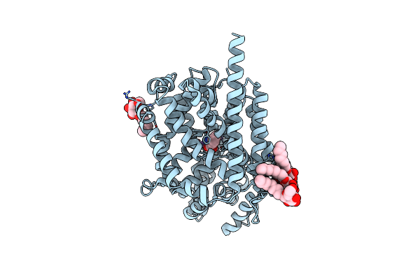 |
Crystal Structure Of Leutaa, A Bacterial Homolog Of Na+/Cl--Dependent Neurotransmitter Transporters
Organism: Aquifex aeolicus
Method: X-RAY DIFFRACTION Resolution:1.65 Å Release Date: 2005-08-02 Classification: TRANSPORT PROTEIN Ligands: BOG, NA, CL, LEU |
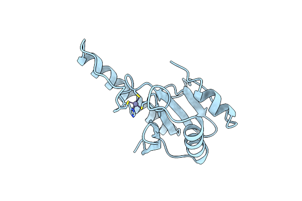 |
Organism: Escherichia coli
Method: X-RAY DIFFRACTION Resolution:1.80 Å Release Date: 1999-10-27 Classification: HYDROLASE Ligands: ZN |

