
Search Count: 8
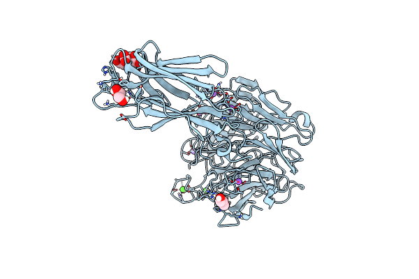 |
Organism: Vibrio cholerae serotype o1
Method: X-RAY DIFFRACTION Resolution:1.90 Å Release Date: 2019-08-28 Classification: SUGAR BINDING PROTEIN Ligands: CA, NA, FLC, GOL |
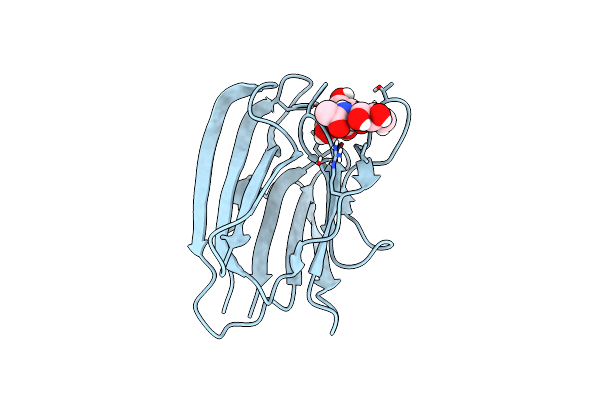 |
Crystal Structure Of The Second Beta-Prism Domain Of Rbmc From V. Cholerae Bound To N-Acetylglucosaminyl-Beta-1,2-Mannose
Organism: Vibrio cholerae o1 biovar el tor str. n16961
Method: X-RAY DIFFRACTION Resolution:1.80 Å Release Date: 2018-01-31 Classification: SUGAR BINDING PROTEIN Ligands: GOL |
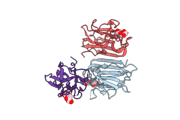 |
Organism: Vibrio cholerae serotype o1 (strain atcc 39315 / el tor inaba n16961)
Method: X-RAY DIFFRACTION Resolution:2.20 Å Release Date: 2018-01-24 Classification: SUGAR BINDING PROTEIN Ligands: GOL |
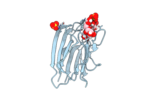 |
Crystal Structure Of The Second Beta-Prism Domain Of Rbmc From V. Cholerae Bound To Mannotriose
Organism: Vibrio cholerae serotype o1 (strain atcc 39315 / el tor inaba n16961)
Method: X-RAY DIFFRACTION Resolution:1.50 Å Release Date: 2018-01-17 Classification: SUGAR BINDING PROTEIN Ligands: SO4 |
 |
 |
Crystal Structure Of The Vibrio Vulnificus Hemolysin/Cytolysin Beta-Trefoil Lectin
Organism: Vibrio vulnificus
Method: X-RAY DIFFRACTION Resolution:2.00 Å Release Date: 2014-05-28 Classification: TOXIN Ligands: GOL |
 |
Crystal Structure Of The Vibrio Vulnificus Hemolysin/Cytolysin Beta-Trefoil Lectin With N-Acetyl-D-Galactosamine Bound
Organism: Vibrio vulnificus
Method: X-RAY DIFFRACTION Resolution:2.00 Å Release Date: 2014-05-28 Classification: TOXIN Ligands: NGA, GOL |
 |
Crystal Structure Of The Vibrio Vulnificus Hemolysin/Cytolysin Beta-Trefoil Lectin With N-Acetyl-D-Lactosamine Bound
Organism: Vibrio vulnificus
Method: X-RAY DIFFRACTION Resolution:2.10 Å Release Date: 2014-05-28 Classification: TOXIN Ligands: GOL, GAL |