
Search Count: 17
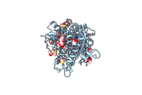 |
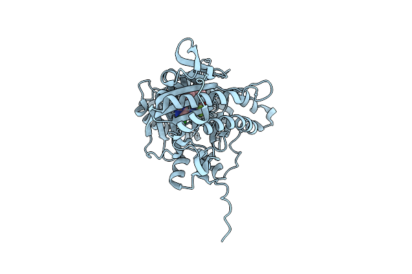
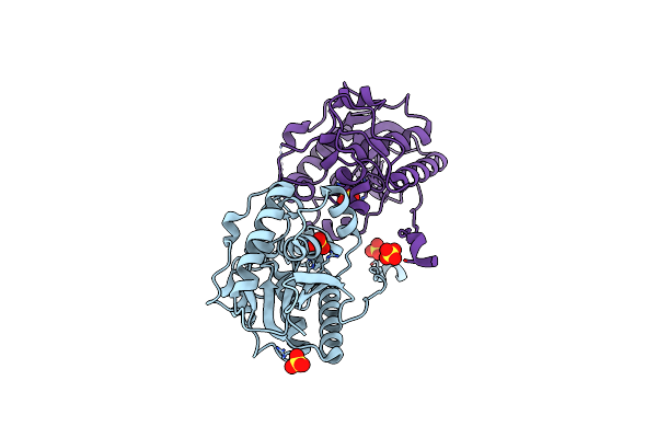
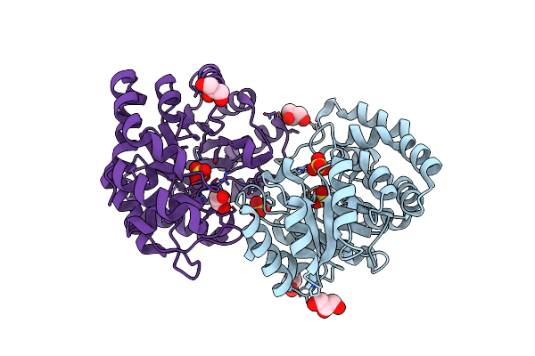
Crystal Structure Of Human Soluble Adenylyl Cyclase (Sac) In Complex With Inhibitor Tdi-09066
Organism: Homo sapiens
Method: X-RAY DIFFRACTION Resolution:1.90 Å Release Date: 2024-01-03 Classification: SIGNALING PROTEIN Ligands: V9E, DMS, ACT, EDO, PG4, PGE, CL |
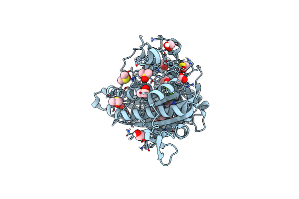 |
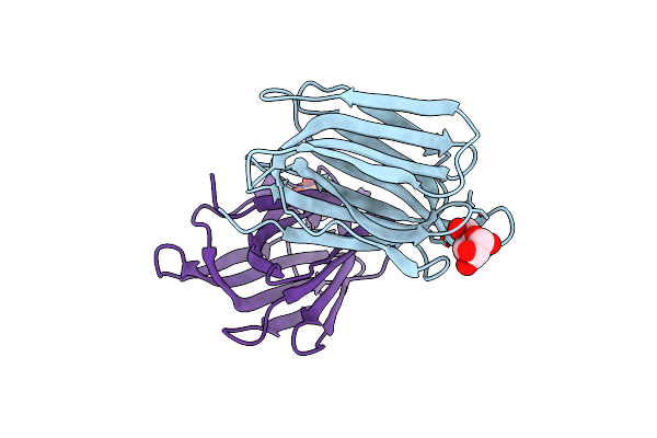
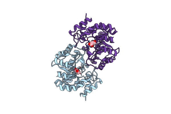
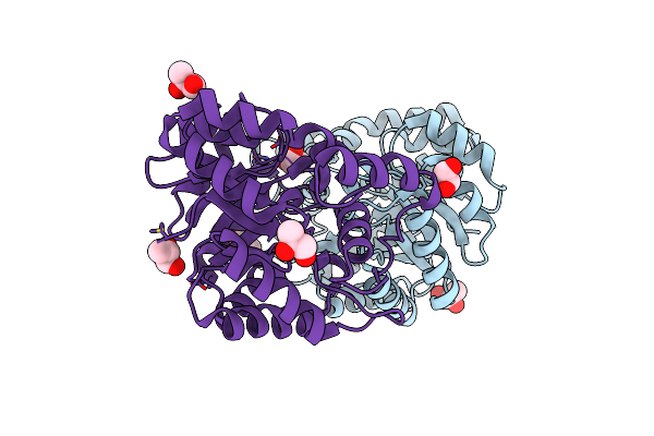
Crystal Structure Of Human Soluble Adenylyl Cyclase (Sac) In Complex With Inhibitor Tdi-10512
Organism: Homo sapiens
Method: X-RAY DIFFRACTION Resolution:2.00 Å Release Date: 2023-04-26 Classification: SIGNALING PROTEIN Ligands: V6U, DMS, EDO, ACT |
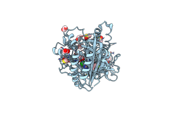 |
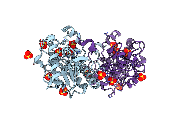
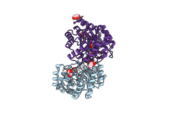
Crystal Structure Of Human Soluble Adenylyl Cyclase Catalytic Domain In Complex With The Inhibitor Tdi-10228
Organism: Homo sapiens
Method: X-RAY DIFFRACTION Resolution:2.10 Å Release Date: 2023-04-26 Classification: SIGNALING PROTEIN Ligands: VE1, DMS, ACT, EDO |
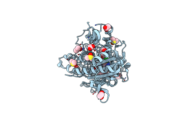 |
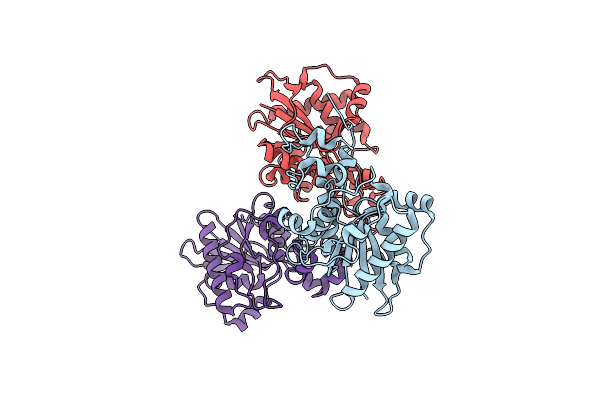
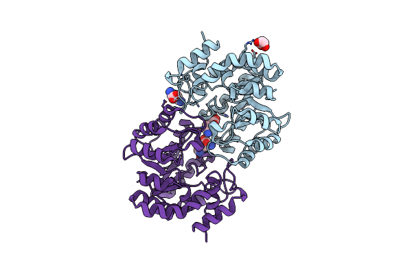
Complex Of Human Soluble Adenylyl Cyclase 10 Catalytic Core With Inhibitor Tdi-10962
Organism: Homo sapiens
Method: X-RAY DIFFRACTION Resolution:2.10 Å Release Date: 2023-04-26 Classification: SIGNALING PROTEIN Ligands: VBB, DMS, EDO, ACT |
 |
Crystal Structure Of Human Soluble Adenylyl Cyclase In Complex With The Inhibitor Tdi-011861
Organism: Homo sapiens
Method: X-RAY DIFFRACTION Resolution:1.82 Å Release Date: 2023-03-29 Classification: SIGNALING PROTEIN Ligands: PJU |
 |
Organism: Homo sapiens
Method: X-RAY DIFFRACTION Resolution:2.20 Å Release Date: 2021-10-06 Classification: SIGNALING PROTEIN Ligands: 1S2, ACT, DMS, EDO |
 |
Organism: Oryza sativa subsp. indica
Method: X-RAY DIFFRACTION Resolution:1.66 Å Release Date: 2017-09-13 Classification: SUGAR BINDING PROTEIN Ligands: MAN |
 |
Organism: Homo sapiens
Method: X-RAY DIFFRACTION Resolution:1.87 Å Release Date: 2017-08-16 Classification: HYDROLASE Ligands: SO4 |
 |
Organism: Homo sapiens
Method: X-RAY DIFFRACTION Resolution:2.48 Å Release Date: 2017-08-16 Classification: HYDROLASE |
 |
Organism: Homo sapiens
Method: X-RAY DIFFRACTION Resolution:2.43 Å Release Date: 2017-08-16 Classification: HYDROLASE Ligands: SO4 |
 |
Structure Of Dihydrodipicolinate Synthase Complexed With 3-Hydroxypropanoic Acid(Hpa)At 2.70 A Resolution
Organism: Pseudomonas aeruginosa
Method: X-RAY DIFFRACTION Resolution:2.70 Å Release Date: 2011-06-15 Classification: LYASE Ligands: 3OH |
 |
Crystal Structure Of The Complex Of Dhydrodipicolinate Synthase From Acinetobacter Baumannii With Lysine At 2.3A Resolution
Organism: Acinetobacter baumannii
Method: X-RAY DIFFRACTION Resolution:2.30 Å Release Date: 2010-12-29 Classification: LYASE Ligands: GOL, SO4, LYS, ACT |
 |
Crystal Structure Of Dihydrodipicolinate Synthase From Pseudomonas Aeruginosa(Psdhdps)Complexed With L-Lysine At 2.65A Resolution
Organism: Pseudomonas aeruginosa
Method: X-RAY DIFFRACTION Resolution:2.65 Å Release Date: 2010-12-29 Classification: LYASE Ligands: LYS, GOL |
 |
Crystal Structure Of Dhydrodipicolinate Synthase From Acinetobacter Baumannii At 2.8A Resolution
Organism: Acinetobacter baumannii
Method: X-RAY DIFFRACTION Resolution:2.80 Å Release Date: 2010-12-22 Classification: LYASE Ligands: SO4, GOL |
 |
Crystal Structure Of The Complex Of Dhydrodipicolinate Synthase From Acinetobacter Baumannii With Lysine At 2.6A Resolution
Organism: Acinetobacter baumannii
Method: X-RAY DIFFRACTION Resolution:2.60 Å Release Date: 2010-12-22 Classification: LYASE Ligands: SO4, LYS, GOL |
 |
Biochemical Studies And Crystal Structure Determination Of Dihydrodipicolinate Synthase From Pseudomonas Aeruginosa
Organism: Pseudomonas aeruginosa
Method: X-RAY DIFFRACTION Resolution:2.85 Å Release Date: 2010-12-15 Classification: LYASE Ligands: PGO |
 |
Crystal Structure Of Dihydrodipicolinate Synthase From Pseudomonas Aeruginosa
Organism: Pseudomonas aeruginosa
Method: X-RAY DIFFRACTION Resolution:2.95 Å Release Date: 2010-07-28 Classification: LYASE Ligands: PGO |