
Search Count: 24
 |
Organism: Streptomyces mobaraensis
Method: X-RAY DIFFRACTION Resolution:2.00 Å Release Date: 2024-09-18 Classification: TRANSFERASE Ligands: MES, CL, PEG |
 |
Crystal Structure Of L-Histidine Decarboxylase (C57S Mutant) From Photobacterium Phosphoreum
Organism: Photobacterium phosphoreum
Method: X-RAY DIFFRACTION Resolution:2.85 Å Release Date: 2022-02-16 Classification: LYASE |
 |
Crystal Structure Of L-Histidine Decarboxylase (C57S/C101V/C282V Mutant) From Photobacterium Phosphoreum
Organism: Photobacterium phosphoreum
Method: X-RAY DIFFRACTION Resolution:2.50 Å Release Date: 2022-02-16 Classification: LYASE Ligands: IMD |
 |
Ambient Temperature Structure Of Bifidobacterium Longum Phosphoketolase With Thiamine Diphosphate
Organism: Bifidobacterium longum
Method: X-RAY DIFFRACTION Resolution:2.50 Å Release Date: 2021-06-02 Classification: LYASE Ligands: TPP, CA, LMR, MLA, SIN |
 |
Ambient Temperature Structure Of Bifidobacgterium Longum Phosphoketolase With Thiamine Diphosphate And Phosphoenol Pyuruvate
Organism: Bifidobacterium longum
Method: X-RAY DIFFRACTION Resolution:2.50 Å Release Date: 2021-06-02 Classification: LYASE Ligands: PEP, TPP, CA |
 |
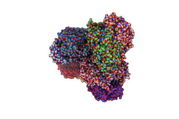
Organism: Bifidobacterium longum subsp. longum f8
Method: ELECTRON MICROSCOPY Release Date: 2021-02-17 Classification: LYASE Ligands: TPP, CA |
 |
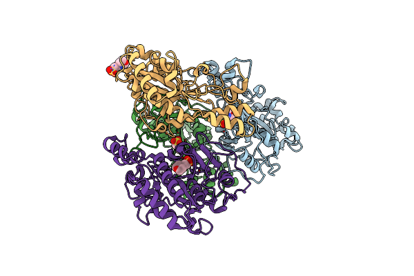
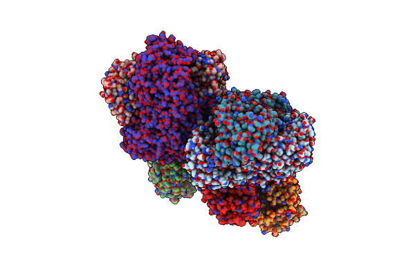
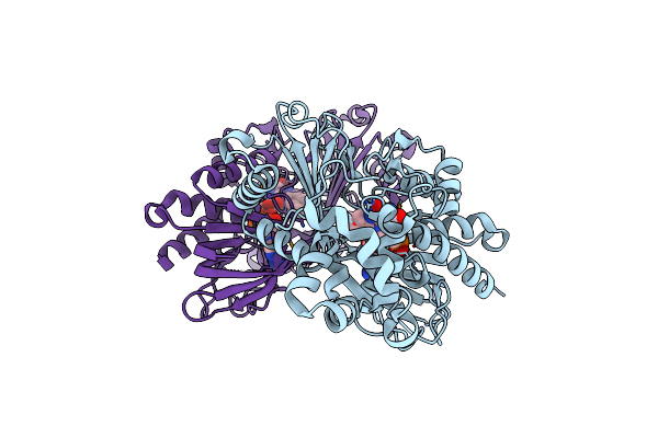
Structure Of Leucine Dehydrogenase From Geobacillus Stearothermophilus By Cryo-Em
Organism: Geobacillus stearothermophilus 10
Method: ELECTRON MICROSCOPY Release Date: 2018-12-26 Classification: OXIDOREDUCTASE |
 |
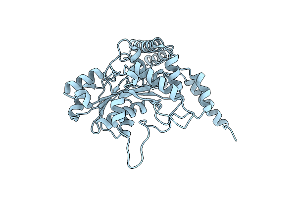
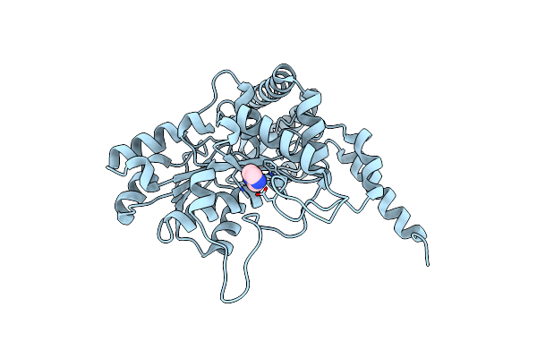
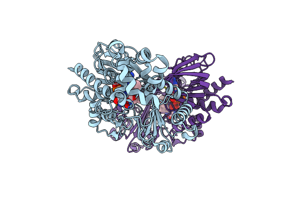
Structure Of Nad+-Bound Leucine Dehydrogenase From Geobacillus Stearothermophilus By Cryo-Em
Organism: Geobacillus stearothermophilus 10
Method: ELECTRON MICROSCOPY Release Date: 2018-12-26 Classification: OXIDOREDUCTASE Ligands: NAD |
 |
Crystal Structure Of Se-Met Tryptophan Oxidase (C395A Mutant) From Chromobacterium Violaceum
Organism: Chromobacterium violaceum (strain atcc 12472 / dsm 30191 / jcm 1249 / nbrc 12614 / ncimb 9131 / nctc 9757)
Method: X-RAY DIFFRACTION Resolution:2.20 Å Release Date: 2018-12-19 Classification: OXIDOREDUCTASE Ligands: FAD |
 |
Crystal Structure Of Tryptophan Oxidase (C395A Mutant) From Chromobacterium Violaceum
Organism: Chromobacterium violaceum (strain atcc 12472 / dsm 30191 / jcm 1249 / nbrc 12614 / ncimb 9131 / nctc 9757)
Method: X-RAY DIFFRACTION Resolution:1.80 Å Release Date: 2018-12-19 Classification: OXIDOREDUCTASE Ligands: TRP, FAD |
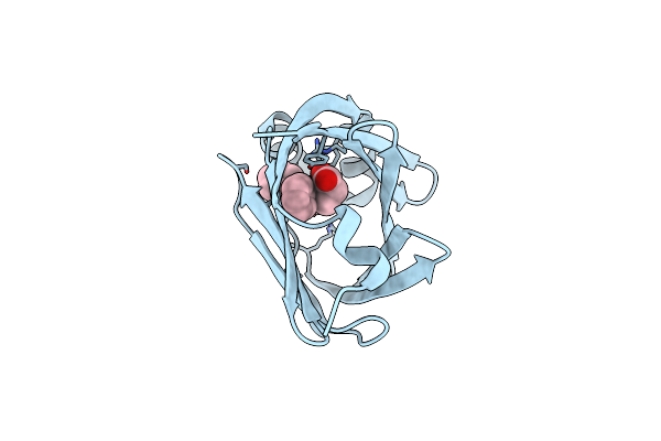 |
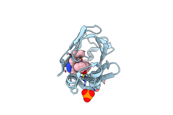
Crystal Structure Of Fabp4 In Complex With 3-(5-Cyclopropyl-2,3-Diphenyl-1H-Indol-1-Yl)Propanoic Acid
Organism: Homo sapiens
Method: X-RAY DIFFRACTION Resolution:1.65 Å Release Date: 2016-06-22 Classification: LIPID BINDING PROTEIN Ligands: 57P |
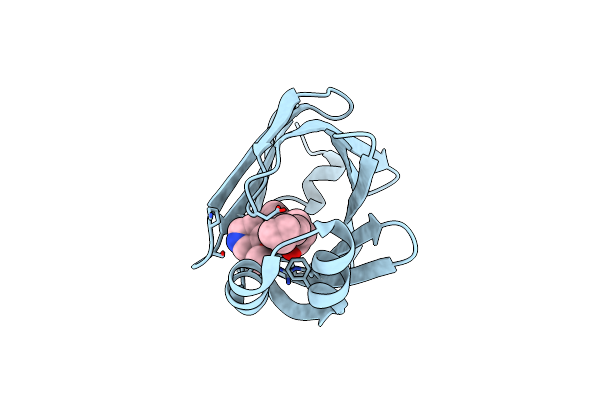 |
Crystal Structure Of Fabp4 In Complex With 3-[5-Cyclopropyl-3-(3-Methoxypyridin-4-Yl)-2-Phenyl-1H-Indol-1-Yl] Propanoic Acid
Organism: Homo sapiens
Method: X-RAY DIFFRACTION Resolution:1.70 Å Release Date: 2016-06-22 Classification: LIPID BINDING PROTEIN Ligands: L19 |
 |
Crystal Structure Of Fabp4 In Complex With 3-{5-Cyclopropyl-3-(3,5-Dimethyl-1H-Pyrazol-4-Yl)-2-[3-(Propan-2-Yloxy) Phenyl]-1H-Indol-1-Yl}Propanoic Acid
Organism: Homo sapiens
Method: X-RAY DIFFRACTION Resolution:1.81 Å Release Date: 2016-06-22 Classification: LIPID BINDING PROTEIN Ligands: L96, PO4 |
 |
Crystal Structure Of Fabp4 In Complex With 3-(2-Phenyl-1H-Indol-1-Yl)Propanoic Acid
Organism: Homo sapiens
Method: X-RAY DIFFRACTION Resolution:1.70 Å Release Date: 2016-06-22 Classification: LIPID BINDING PROTEIN Ligands: 57Q |
 |
Organism: Bifidobacterium longum
Method: X-RAY DIFFRACTION Resolution:2.20 Å Release Date: 2010-09-15 Classification: LYASE Ligands: TPP, CA |
 |
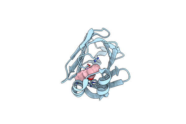
Organism: Curculigo latifolia
Method: X-RAY DIFFRACTION Resolution:1.50 Å Release Date: 2007-05-15 Classification: PLANT PROTEIN Ligands: SO4 |
 |
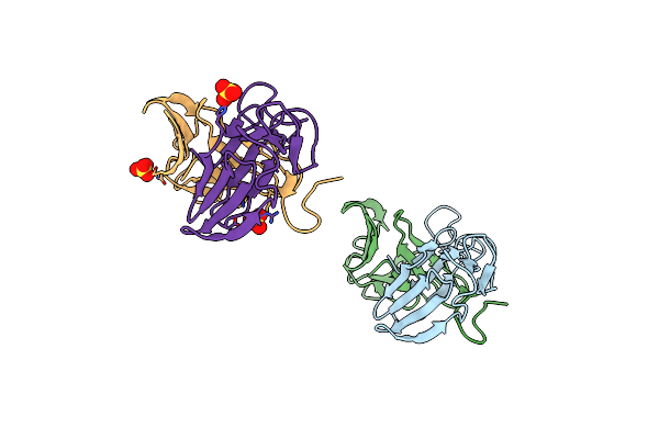
Organism: Pseudomonas aeruginosa
Method: X-RAY DIFFRACTION Resolution:2.56 Å Release Date: 2004-11-02 Classification: MEMBRANE PROTEIN |
 |
Organism: Streptomyces mobaraensis
Method: X-RAY DIFFRACTION Resolution:2.40 Å Release Date: 2002-08-27 Classification: TRANSFERASE |
 |
Organism: Pichia farinosa
Method: X-RAY DIFFRACTION Resolution:1.80 Å Release Date: 1997-04-01 Classification: TOXIN Ligands: SO4 |
 |
Organism: Pichia farinosa
Method: X-RAY DIFFRACTION Resolution:1.80 Å Release Date: 1997-04-01 Classification: TOXIN |