
Search Count: 18
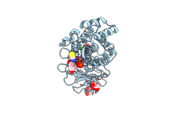 |
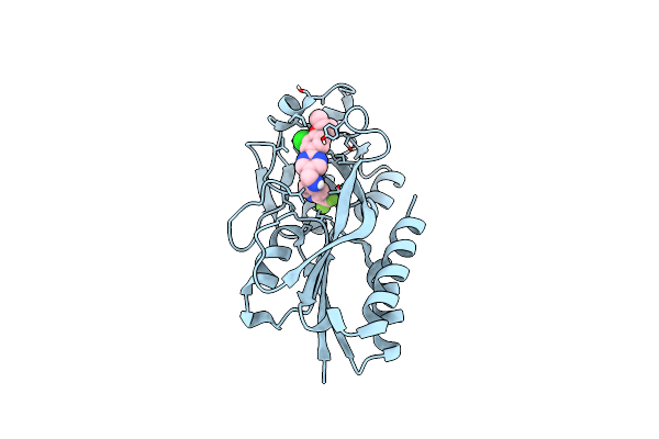
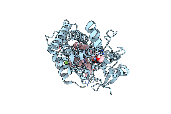
Crystal Structure Of Recombinant Lasb From Pseudomonas Aeruginosa Pao1 In Complex With 6466
Organism: Pseudomonas aeruginosa pao1
Method: X-RAY DIFFRACTION Resolution:1.31 Å Release Date: 2024-10-16 Classification: HYDROLASE Ligands: ZN, CA, GOL, SCN, XI5 |
 |
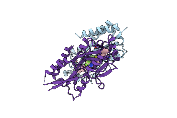
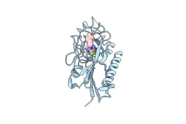
Organism: Pseudomonas aeruginosa
Method: X-RAY DIFFRACTION Resolution:2.70 Å Release Date: 2023-12-06 Classification: HYDROLASE Ligands: CA, U7F, ZN |
 |
Crystal Structure Of Pqsr (Mvfr) Ligand-Binding Domain In Complex With Compound N-((2-(4-Cyclopropylphenyl)Thiazol-5-Yl)Methyl)-2-(Trifluoromethyl)Pyridin-4-Amine
Organism: Pseudomonas aeruginosa (strain atcc 15692 / dsm 22644 / cip 104116 / jcm 14847 / lmg 12228 / 1c / prs 101 / pao1)
Method: X-RAY DIFFRACTION Resolution:2.65 Å Release Date: 2022-11-30 Classification: GENE REGULATION Ligands: XBH |
 |
Crystal Structure Of Pqsr (Mvfr) Ligand-Binding Domain In Complex With Compound 1456
Organism: Pseudomonas aeruginosa (strain atcc 15692 / dsm 22644 / cip 104116 / jcm 14847 / lmg 12228 / 1c / prs 101 / pao1)
Method: X-RAY DIFFRACTION Resolution:2.67 Å Release Date: 2022-11-23 Classification: GENE REGULATION Ligands: 9ZL |
 |
Crystal Structure Of Pqsr (Mvfr) Ligand-Binding Domain In Complex With Compound N-((2-(4-Phenoxyphenyl)Thiazol-5-Yl)Methyl)-2-(Trifluoromethyl)Pyridin-4-Amine
Organism: Pseudomonas aeruginosa (strain atcc 15692 / dsm 22644 / cip 104116 / jcm 14847 / lmg 12228 / 1c / prs 101 / pao1)
Method: X-RAY DIFFRACTION Resolution:2.67 Å Release Date: 2022-11-23 Classification: GENE REGULATION Ligands: A0F |
 |
Crystal Structure Of Pqsr (Mvfr) Ligand-Binding Domain In Complex With 3-Pyridin-4-Yl-2,4-Dihydro-Indeno[1,2-.C.]Pyrazole
Organism: Pseudomonas aeruginosa (strain atcc 15692 / dsm 22644 / cip 104116 / jcm 14847 / lmg 12228 / 1c / prs 101 / pao1)
Method: X-RAY DIFFRACTION Resolution:2.74 Å Release Date: 2022-07-27 Classification: GENE REGULATION Ligands: 5N9 |
 |
Organism: Pseudomonas aeruginosa
Method: X-RAY DIFFRACTION Resolution:1.95 Å Release Date: 2022-01-19 Classification: HYDROLASE Ligands: ZN, V85, CA |
 |
Crystal Structure Of Pqsr (Mvfr) Ligand-Binding Domain In Complex With A Pyridin Agonist
Organism: Pseudomonas aeruginosa pao1
Method: X-RAY DIFFRACTION Resolution:2.28 Å Release Date: 2021-10-06 Classification: DNA BINDING PROTEIN Ligands: U7Q |
 |
Organism: Mycobacterium tuberculosis
Method: X-RAY DIFFRACTION Resolution:1.50 Å Release Date: 2021-05-26 Classification: OXIDOREDUCTASE Ligands: N55, PGE, EDO, HEM |
 |
Organism: Mycobacterium tuberculosis
Method: X-RAY DIFFRACTION Resolution:1.50 Å Release Date: 2021-05-26 Classification: OXIDOREDUCTASE Ligands: EDO, N5Z, PGE, HEM |
 |
Organism: Mycobacterium tuberculosis
Method: X-RAY DIFFRACTION Resolution:1.70 Å Release Date: 2021-05-26 Classification: OXIDOREDUCTASE Ligands: N5W, EDO, HEM |
 |
Crystal Structure Of Pqsr (Mvfr) Ligand-Binding Domain In Complex With Triazolo-Pyridine Inverse Agonist A
Organism: Pseudomonas aeruginosa (strain atcc 15692 / dsm 22644 / cip 104116 / jcm 14847 / lmg 12228 / 1c / prs 101 / pao1)
Method: X-RAY DIFFRACTION Resolution:2.16 Å Release Date: 2021-04-14 Classification: DNA BINDING PROTEIN Ligands: OT2, OT8, MG |
 |
Organism: Pseudomonas aeruginosa pao1
Method: X-RAY DIFFRACTION Resolution:3.14 Å Release Date: 2019-11-20 Classification: DNA BINDING PROTEIN Ligands: HLH |
 |
Crystal Structure Of Pqsr (Mvfr) Ligand-Binding Domain In Complex With Compound 11
Organism: Pseudomonas aeruginosa pao1
Method: X-RAY DIFFRACTION Resolution:2.56 Å Release Date: 2019-11-20 Classification: DNA BINDING PROTEIN Ligands: HLK |
 |
Crystal Structure Of Pqsr (Mvfr) Ligand-Binding Domain In Complex With Compound 20
Organism: Pseudomonas aeruginosa pao1
Method: X-RAY DIFFRACTION Resolution:2.82 Å Release Date: 2019-11-20 Classification: DNA BINDING PROTEIN Ligands: HLQ, GOL |
 |
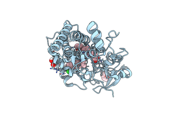
Organism: Pseudomonas aeruginosa
Method: X-RAY DIFFRACTION Resolution:2.10 Å Release Date: 2018-09-05 Classification: HYDROLASE Ligands: EEK, ZN, CA, SO4, GOL |
 |
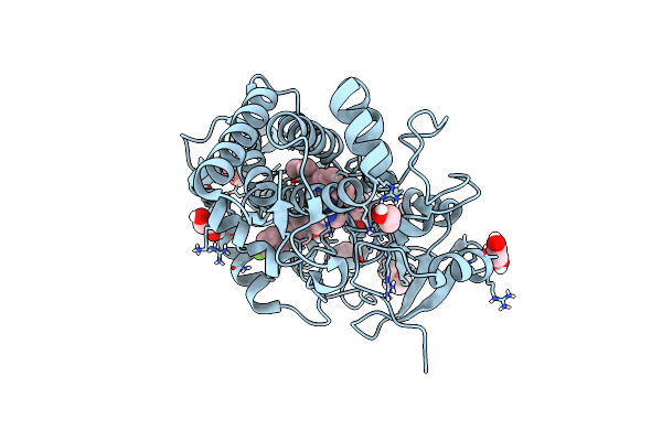
Organism: Pseudomonas aeruginosa
Method: X-RAY DIFFRACTION Resolution:1.30 Å Release Date: 2018-03-28 Classification: HYDROLASE Ligands: CA, ZN, CXH |
 |
Crystal Structure Of The Peptidase Domain Of Collagenase H From Clostridium Histolyticum In Complex With N-Aryl Mercaptoacetamide-Based Inhibitor
Organism: Hathewaya histolytica
Method: X-RAY DIFFRACTION Resolution:1.87 Å Release Date: 2018-01-31 Classification: HYDROLASE Ligands: ZN, CA, 9NB |