
Search Count: 152
 |
Organism: Pyrococcus horikoshii ot3
Method: X-RAY DIFFRACTION Resolution:3.10 Å Release Date: 2025-03-12 Classification: TRANSFERASE Ligands: SO4, SO3, EDO, PEG |
 |
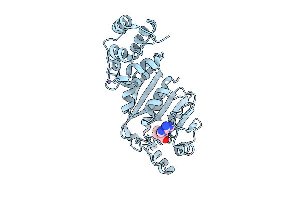
Crystal Structure Of Nep1 In Complex With Adenosine From Pyrococcus Horikoshii Ot3
Organism: Pyrococcus horikoshii ot3
Method: X-RAY DIFFRACTION Resolution:2.01 Å Release Date: 2025-03-12 Classification: TRANSFERASE Ligands: CL, ADN, SO4, SO3, EDO, PEG |
 |
Crystal Structure Of Nep1 In Complex With 5'-Methylthioadenosine From Pyrococcus Horikoshii Ot3
Organism: Pyrococcus horikoshii ot3
Method: X-RAY DIFFRACTION Resolution:2.20 Å Release Date: 2025-03-12 Classification: TRANSFERASE Ligands: CL, MTA, EDO, GOL, ACT |
 |
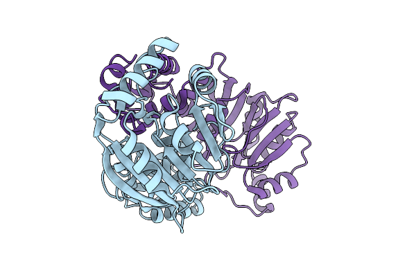
Crystal Structure Of Rrna (Uracil-C5)-Methyltransferase From Pyrococcus Horikoshii Ot3
Organism: Pyrococcus horikoshii ot3
Method: X-RAY DIFFRACTION Resolution:3.20 Å Release Date: 2024-10-16 Classification: TRANSFERASE |
 |
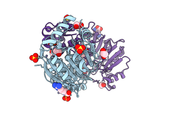
Crystal Structure Of Dimt1 From The Thermophilic Archaeon, Pyrococcus Horikoshii
Organism: Pyrococcus horikoshii ot3
Method: X-RAY DIFFRACTION Resolution:2.01 Å Release Date: 2024-08-28 Classification: TRANSFERASE Ligands: ZN, SO3, GOL, PEG, EDO, ACT, ARG, EPE |
 |
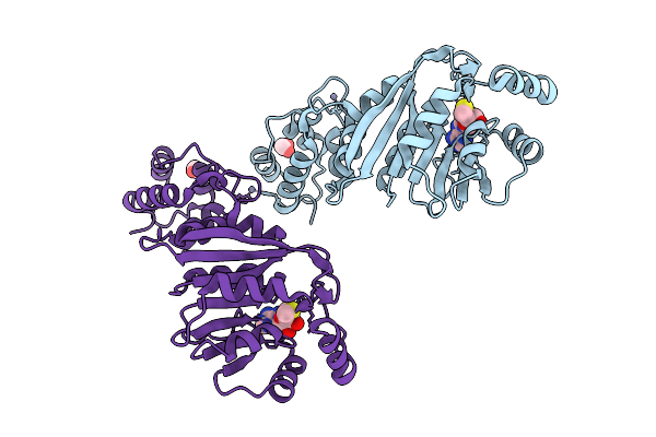
Crystal Structure Of Dimt1 In Complex With 5'-Methylthioadenosine And Adenosine From Pyrococcus Horikoshii
Organism: Pyrococcus horikoshii ot3
Method: X-RAY DIFFRACTION Resolution:2.35 Å Release Date: 2024-08-28 Classification: TRANSFERASE Ligands: MTA, ADN, ZN, CL, SO3, GOL, EDO, ACT, ARG, PEG |
 |
Crystal Structure Of Dimt1 In Complex With 5'-Methylthioadenosine From Pyrococcus Horikoshii (Formi)
Organism: Pyrococcus horikoshii ot3
Method: X-RAY DIFFRACTION Resolution:2.60 Å Release Date: 2024-08-28 Classification: TRANSFERASE Ligands: MTA, ZN, SO3, PEG, CL, GOL, EDO |
 |
Crystal Structure Of Dimt1 In Complex With 5'-Methylthioadenosine From Pyrococcus Horikoshii (Formii)
Organism: Pyrococcus horikoshii ot3
Method: X-RAY DIFFRACTION Resolution:2.50 Å Release Date: 2024-08-28 Classification: TRANSFERASE Ligands: MTA, ZN, SO3, GOL, EDO, PEG, PGE, ACT |
 |
Crystal Structure Of Dimt1 In Complex With Adenosylornithine (Sfg) From Pyrococcus Horikoshii
Organism: Pyrococcus horikoshii ot3
Method: X-RAY DIFFRACTION Resolution:2.20 Å Release Date: 2024-08-28 Classification: TRANSFERASE Ligands: SFG, ZN, CL, PEG, ARG, SO3, EDO |
 |
Crystal Structure Of Dimt1 In Complex With S-Adenosyl-L-Homocysteine (Sah) From Pyrococcus Horikoshii
Organism: Pyrococcus horikoshii ot3
Method: X-RAY DIFFRACTION Resolution:2.80 Å Release Date: 2024-08-28 Classification: TRANSFERASE Ligands: SAH, ZN, SO3, EDO, GOL, PEG, PGE, ARG |
 |
Organism: Pyrococcus horikoshii ot3
Method: X-RAY DIFFRACTION Resolution:3.30 Å Release Date: 2024-08-28 Classification: TRANSFERASE |
 |
Crystal Structure Of The D117A Mutant Of Dimt1 In Complex With 5'-Methylthioadenosine From Pyrococcus Horikoshii
Organism: Pyrococcus horikoshii ot3
Method: X-RAY DIFFRACTION Resolution:3.30 Å Release Date: 2024-08-28 Classification: TRANSFERASE Ligands: MTA, ZN |
 |
Organism: Pyrococcus horikoshii ot3
Method: X-RAY DIFFRACTION Resolution:2.60 Å Release Date: 2024-08-28 Classification: TRANSFERASE Ligands: ZN, SO3, EDO, ARG, GOL, PEG |
 |
Crystal Structure Of The Y135A Mutant Of Dimt1 In Complex With 5'-Methylthioadenosine From Pyrococcus Horikoshii
Organism: Pyrococcus horikoshii ot3
Method: X-RAY DIFFRACTION Resolution:3.00 Å Release Date: 2024-08-28 Classification: TRANSFERASE Ligands: MTA, ZN, EDO |
 |
Crystal Structure Of The Y135A Mutant Of Dimt1 In Complex With Adenosylornithine (Sfg) From Pyrococcus Horikoshii
Organism: Pyrococcus horikoshii ot3
Method: X-RAY DIFFRACTION Resolution:2.60 Å Release Date: 2024-08-28 Classification: TRANSFERASE Ligands: ZN, SFG |
 |
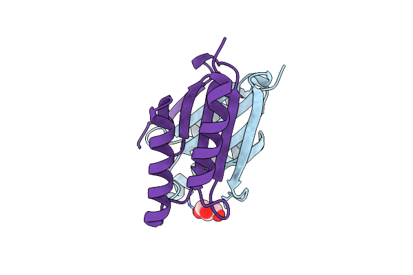
Crystal Structure Of The Mlad Domain Of The Mlad Protein From Escherichia Coli (Form I)
Organism: Escherichia coli k-12
Method: X-RAY DIFFRACTION Resolution:2.30 Å Release Date: 2024-01-10 Classification: TRANSPORT PROTEIN Ligands: CO2, EDO |
 |
Crystal Structure Of The Mlad Domain Of The Mlad Protein From Escherichia Coli (Form Ii)
Organism: Escherichia coli k-12
Method: X-RAY DIFFRACTION Resolution:2.70 Å Release Date: 2024-01-10 Classification: TRANSPORT PROTEIN Ligands: MG, CO2 |
 |
Crystal Structure Of The Ectodomain Of The Mlad Protein From Escherichia Coli In The Resting State
Organism: Escherichia coli k-12
Method: X-RAY DIFFRACTION Resolution:3.20 Å Release Date: 2024-01-10 Classification: TRANSPORT PROTEIN |
 |
Organism: Leptospira interrogans serovar copenhageni str. fiocruz l1-130
Method: X-RAY DIFFRACTION Resolution:2.60 Å Release Date: 2022-07-06 Classification: DNA BINDING PROTEIN Ligands: GOL |
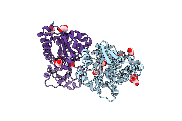 |
Crystal Structure Of An Orphan Heme Uptake Protein (Mhup) Of Abc Transporter From Mycobacterium Tuberculosis (Form I)
Organism: Mycobacterium tuberculosis h37rv
Method: X-RAY DIFFRACTION Resolution:1.80 Å Release Date: 2022-06-01 Classification: TRANSPORT PROTEIN Ligands: CIT, EDO, GOL, CO2, CO3, ACT |