
Search Count: 36
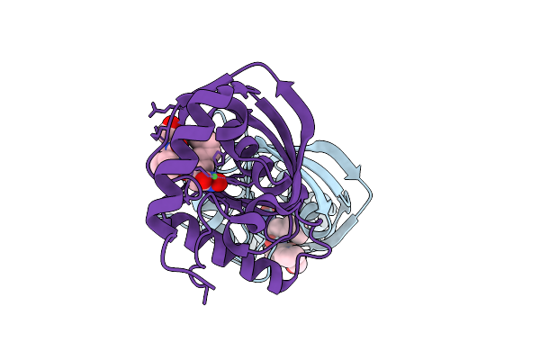 |
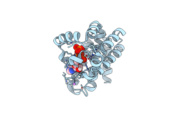
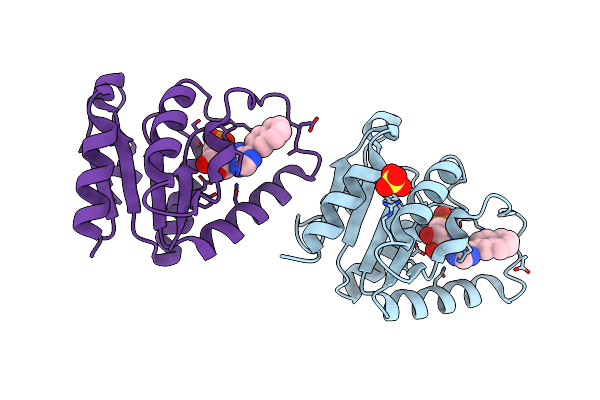
Organism: Vibrio cholerae o1 biovar el tor str. n16961
Method: X-RAY DIFFRACTION Release Date: 2025-12-10 Classification: HYDROLASE Ligands: NI, BB2 |
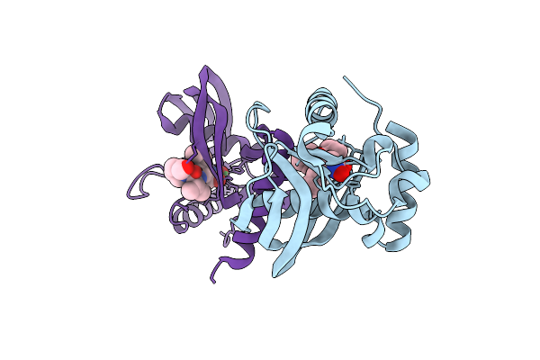 |
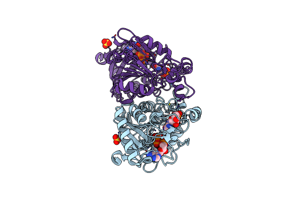
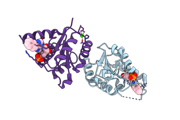
Organism: Vibrio cholerae o1 biovar el tor str. n16961
Method: X-RAY DIFFRACTION Release Date: 2025-12-10 Classification: HYDROLASE Ligands: BB2, NI |
 |
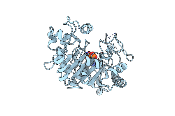
Crystal Structure Of A 2`-Deoxyribosyltransferase From The Psychrophilic Bacterium Desulfotalea Psychrophila.
Organism: Desulfotalea psychrophila (strain lsv54 / dsm 12343)
Method: X-RAY DIFFRACTION Resolution:2.40 Å Release Date: 2021-10-20 Classification: TRANSFERASE Ligands: GOL |
 |
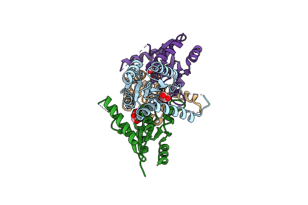
Cyanophage S-2L Succinoaminodeoxyadenylate Synthetase (Purz) Bound To Dgmp And Datp As An Energy Donor
Organism: Cyanophage s-2l
Method: X-RAY DIFFRACTION Resolution:1.70 Å Release Date: 2021-09-01 Classification: VIRAL PROTEIN Ligands: DGP, DTP |
 |
Cyanophage S-2L Mazg-Like Pyrophosphohydrolase Bound To Dgdp And Three Catalytic Mn2+ Ions Per Active Site
Organism: Cyanophage s-2l
Method: X-RAY DIFFRACTION Resolution:1.43 Å Release Date: 2021-09-01 Classification: VIRAL PROTEIN Ligands: DGI, MN, SO4 |
 |
Organism: Streptococcus agalactiae
Method: X-RAY DIFFRACTION Resolution:2.64 Å Release Date: 2021-08-18 Classification: SIGNALING PROTEIN Ligands: SO4, PT |
 |
Organism: Streptococcus agalactiae
Method: X-RAY DIFFRACTION Release Date: 2021-08-18 Classification: SIGNALING PROTEIN Ligands: 2BA, SO4 |
 |
Organism: Cyanophage s-2l
Method: X-RAY DIFFRACTION Resolution:1.50 Å Release Date: 2021-03-03 Classification: VIRAL PROTEIN Ligands: CA |
 |
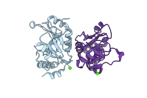
Cyanophage S-2L Hd Phosphohydrolase (Datz) Bound To Da And One Catalytic Zn2+ Ion
Organism: Cyanophage s-2l
Method: X-RAY DIFFRACTION Resolution:0.86 Å Release Date: 2021-03-03 Classification: VIRAL PROTEIN Ligands: 3D1, ZN, LI |
 |
Cyanophage S-2L Hd Phosphohydrolase (Datz) Bound To Da And Two Catalytic Co2+ Ions
Organism: Cyanophage s-2l
Method: X-RAY DIFFRACTION Resolution:1.72 Å Release Date: 2021-03-03 Classification: VIRAL PROTEIN Ligands: 3D1, CO |
 |
Organism: Cyanophage s-2l
Method: X-RAY DIFFRACTION Resolution:1.27 Å Release Date: 2021-03-03 Classification: VIRAL PROTEIN Ligands: DTP, ZN, LI |
 |
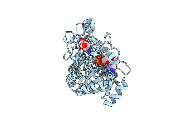
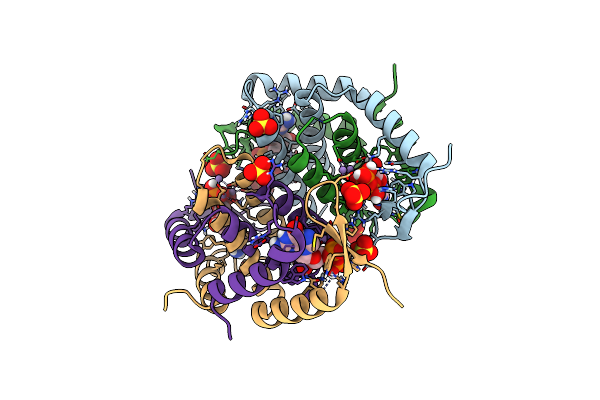
Deoxyguanylosuccinate Synthase (Dgss) Quaternary Structure With Amppcp, Dgmp, Asp, Magnesium At 2.21 Angstrom Resolution
Organism: Vibrio phage phivc8
Method: X-RAY DIFFRACTION Resolution:2.21 Å Release Date: 2020-12-16 Classification: BIOSYNTHETIC PROTEIN Ligands: SO4, DGP, ACP, ASP, MG |
 |
Organism: Vibrio phage phivc8
Method: X-RAY DIFFRACTION Resolution:2.50 Å Release Date: 2020-06-03 Classification: BIOSYNTHETIC PROTEIN Ligands: ATP, MG, IMP, SO4 |
 |
Deoxyguanylosuccinate Synthase (Dgss) Quaternary Structure With Atp, Dgmp, Hadacidin At 2.1 Angstrom Resolution
Organism: Vibrio phage phivc8
Method: X-RAY DIFFRACTION Resolution:2.10 Å Release Date: 2019-06-26 Classification: BIOSYNTHETIC PROTEIN Ligands: HDA, DGP, FLC, ATP |
 |
Deoxyguanylosuccinate Synthase (Dgss) Structure At 1.33 Angstrom Resolution.
Organism: Vibrio phage phivc8
Method: X-RAY DIFFRACTION Resolution:1.33 Å Release Date: 2019-06-12 Classification: BIOSYNTHETIC PROTEIN |
 |
Deoxyguanylosuccinate Synthase (Dgss) And Atp Structure At 1.7 Angstrom Resolution
Organism: Vibrio phage phivc8
Method: X-RAY DIFFRACTION Resolution:1.70 Å Release Date: 2019-06-12 Classification: BIOSYNTHETIC PROTEIN Ligands: ATP |
 |
Deoxyguanylosuccinate Synthase (Dgss) Quaternary Structure With Atpanddgmp At 2.3 Angstrom Resolution
Organism: Vibrio phage phivc8
Method: X-RAY DIFFRACTION Resolution:2.35 Å Release Date: 2019-06-12 Classification: BIOSYNTHETIC PROTEIN Ligands: ATP, DGP, MG, CL, GOL |
 |
Deoxyguanylosuccinate Synthase (Dgss) Structure With Adp At 1.9 Angstrom Resolution
Organism: Vibrio phage phivc8
Method: X-RAY DIFFRACTION Resolution:1.95 Å Release Date: 2019-06-12 Classification: BIOSYNTHETIC PROTEIN Ligands: ADP, CL |
 |
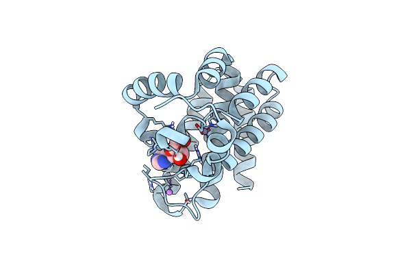
Crystal Structure Of Rat Dnph1 (Rcl) With 6-Naphthyl-Purine-Riboside-Monophosphate
Organism: Rattus norvegicus
Method: X-RAY DIFFRACTION Resolution:2.11 Å Release Date: 2014-08-20 Classification: HYDROLASE Ligands: N6P, SO4 |
 |
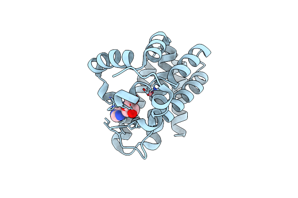
Crystal Structure Of Human Dnph1 (Rcl) With 6-Naphthyl-Purine-Riboside-Monophosphate
Organism: Homo sapiens
Method: X-RAY DIFFRACTION Resolution:1.35 Å Release Date: 2014-08-20 Classification: HYDROLASE Ligands: N6P, CA |