
Search Count: 117
 |
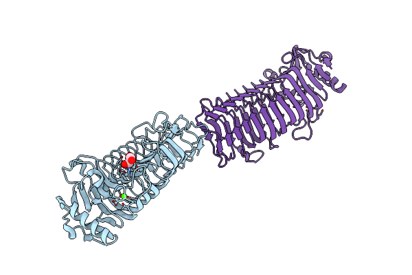
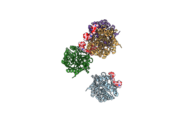
Crystal Structure Of The Gracilariopsis Lemaneiformis Alpha-1,4- Glucan Lyase Covalent Intermediate Complex With 5-Fluoro-Idosyl- Fluoride
Organism: Gracilariopsis lemaneiformis
Method: X-RAY DIFFRACTION Resolution:1.90 Å Release Date: 2013-03-27 Classification: LYASE Ligands: B9D, GOL, 5DI |
 |
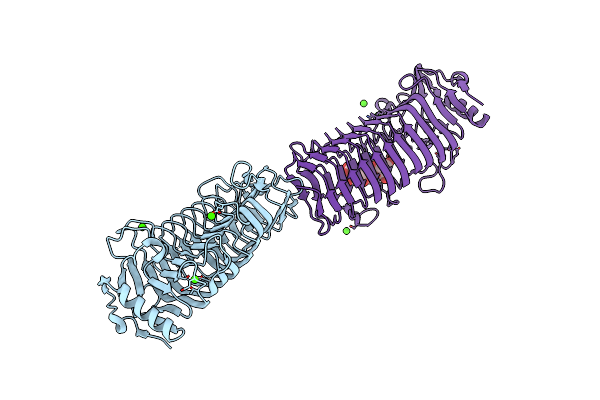
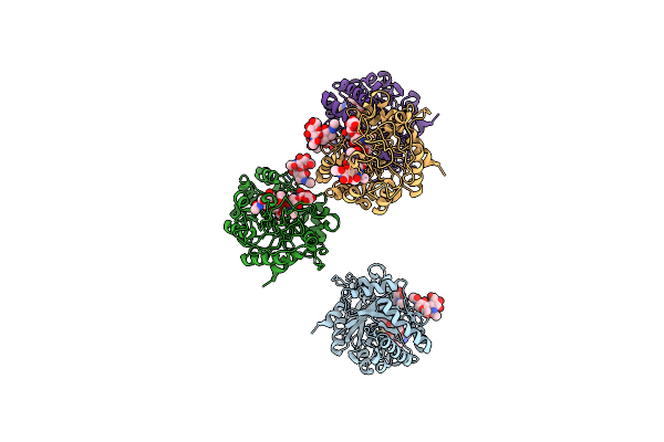
Crystal Structure Of The Gracilariopsis Lemaneiformis Alpha-1,4- Glucan Lyase Covalent Intermediate Complex With 5-Fluoro-Glucosyl- Fluoride
Organism: Gracilariopsis lemaneiformis
Method: X-RAY DIFFRACTION Resolution:2.10 Å Release Date: 2013-03-27 Classification: LYASE Ligands: 5GF, GOL, AFR |
 |
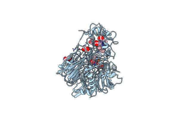
Crystal Structure Of The Gracilariopsis Lemaneiformis Alpha-1,4- Glucan Lyase
Organism: Gracilariopsis lemaneiformis
Method: X-RAY DIFFRACTION Resolution:2.06 Å Release Date: 2011-01-19 Classification: LYASE Ligands: GOL, ACT, CL |
 |
Crystal Structure Of The Gracilariopsis Lemaneiformis Alpha-1,4- Glucan Lyase With Acarbose
Organism: Gracilariopsis lemaneiformis
Method: X-RAY DIFFRACTION Resolution:2.60 Å Release Date: 2011-01-19 Classification: LYASE Ligands: GOL |
 |
Crystal Structure Of The Gracilariopsis Lemaneiformis Alpha- 1,4-Glucan Lyase With Deoxynojirimycin
Organism: Gracilariopsis lemaneiformis
Method: X-RAY DIFFRACTION Resolution:2.35 Å Release Date: 2011-01-19 Classification: LYASE Ligands: NOJ, GOL, CL |
 |
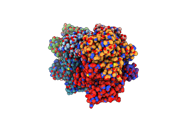
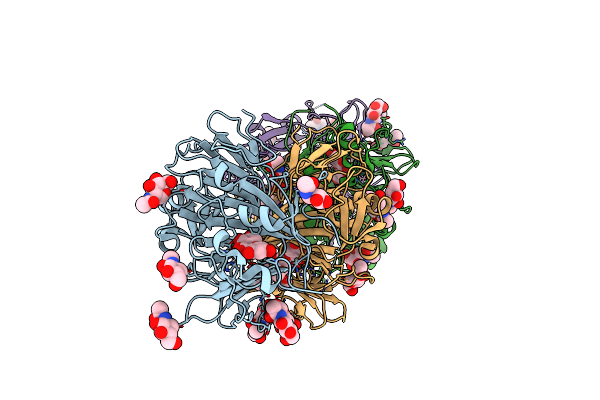
Structure Of Glycosomal Glyceraldehyde-3-Phosphate Dehydrogenase From Trypanosoma Brucei Determined From Laue Data
Organism: Trypanosoma brucei brucei
Method: X-RAY DIFFRACTION Resolution:3.20 Å Release Date: 2009-12-22 Classification: OXIDOREDUCTASE Ligands: SO4, NAD |
 |
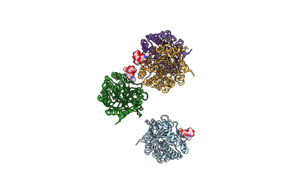
Organism: Azotobacter vinelandii
Method: X-RAY DIFFRACTION Resolution:2.10 Å Release Date: 2008-05-27 Classification: ISOMERASE Ligands: CA, GOL |
 |
Azotobacter Vinelandii Mannuronan C-5 Epimerase Alge4 A-Module Complexed With Mannuronan Trisaccharide
Organism: Azotobacter vinelandii
Method: X-RAY DIFFRACTION Resolution:2.70 Å Release Date: 2008-05-27 Classification: ISOMERASE Ligands: CA |
 |
Organism: Arthrobacter sp. ad2
Method: X-RAY DIFFRACTION Resolution:2.00 Å Release Date: 2006-04-25 Classification: LYASE |
 |
Crystal Structure Of The Haloalcohol Dehalogenase Hhec Complexed With Bromide
Organism: Agrobacterium tumefaciens
Method: X-RAY DIFFRACTION Resolution:1.80 Å Release Date: 2003-10-07 Classification: LYASE Ligands: BR |
 |
Crystal Structure Of The Haloalcohol Dehalogenase Hhec Complexed With (R)-Styrene Oxide And Chloride
Organism: Agrobacterium tumefaciens
Method: X-RAY DIFFRACTION Resolution:2.50 Å Release Date: 2003-10-07 Classification: LYASE Ligands: CL, RSO |
 |
Crystal Structure Of The Haloalcohol Dehalogenase Hhec Complexed With The Haloalcohol Mimic (R)-1-Para-Nitro-Phenyl-2-Azido-Ethanol
Organism: Agrobacterium tumefaciens
Method: X-RAY DIFFRACTION Resolution:1.90 Å Release Date: 2003-10-07 Classification: LYASE Ligands: RPN |
 |
Organism: Homo sapiens
Method: X-RAY DIFFRACTION Resolution:2.70 Å Release Date: 2003-08-26 Classification: SIGNALING PROTEIN |
 |
Crystal Structure Of Human Cartilage Gp39 (Hc-Gp39) In Complex With Chitobiose
Organism: Homo sapiens
Method: X-RAY DIFFRACTION Resolution:2.70 Å Release Date: 2003-08-26 Classification: SIGNALING PROTEIN |
 |
Crystal Structure Of Human Cartilage Gp39 (Hc-Gp39) In Complex With Chitopentaose
Organism: Homo sapiens
Method: X-RAY DIFFRACTION Resolution:2.50 Å Release Date: 2003-08-26 Classification: SIGNALING PROTEIN |
 |
Crystal Structure Of Human Cartilage Gp39 (Hc-Gp39) In Complex With Chitotetraose
Organism: Homo sapiens
Method: X-RAY DIFFRACTION Resolution:2.20 Å Release Date: 2003-08-26 Classification: SIGNALING PROTEIN |
 |
Crystal Structure Of Quercetin 2,3-Dioxygenase Anaerobically Complexed With The Substrate Quercetn
Organism: Aspergillus japonicus
Method: X-RAY DIFFRACTION Resolution:1.75 Å Release Date: 2002-11-28 Classification: OXIDOREDUCTASE Ligands: NAG, CU, QUE, MPD |
 |
Crystal Structure Of Quercetin 2,3-Dioxygenase Anaerobically Complexed With The Substrate Kaempferol
Organism: Aspergillus japonicus
Method: X-RAY DIFFRACTION Resolution:1.90 Å Release Date: 2002-11-28 Classification: OXIDOREDUCTASE Ligands: NAG, KMP, CU, MPD |
 |
Organism: Aspergillus japonicus
Method: X-RAY DIFFRACTION Resolution:1.60 Å Release Date: 2002-05-22 Classification: OXIDOREDUCTASE Ligands: NAG, CU, EDO |
 |
Crystal Structure Of Quinohemoprotein Alcohol Dehydrogenase From Comamonas Testosteroni
Organism: Comamonas testosteroni
Method: X-RAY DIFFRACTION Resolution:1.44 Å Release Date: 2001-12-28 Classification: OXIDOREDUCTASE Ligands: CA, HEC, TFB, PQQ, GOL |