
Search Count: 17
 |
Organism: Uncultured gammaproteobacteria bacterium
Method: ELECTRON MICROSCOPY Release Date: 2024-07-03 Classification: PROTON TRANSPORT Ligands: DKA |
 |
Organism: Uncultured gammaproteobacteria bacterium
Method: ELECTRON MICROSCOPY Release Date: 2024-07-03 Classification: PROTON TRANSPORT Ligands: RET |
 |
Organism: Uncultured gammaproteobacteria bacterium
Method: ELECTRON MICROSCOPY Release Date: 2024-07-03 Classification: PROTON TRANSPORT Ligands: RET |
 |
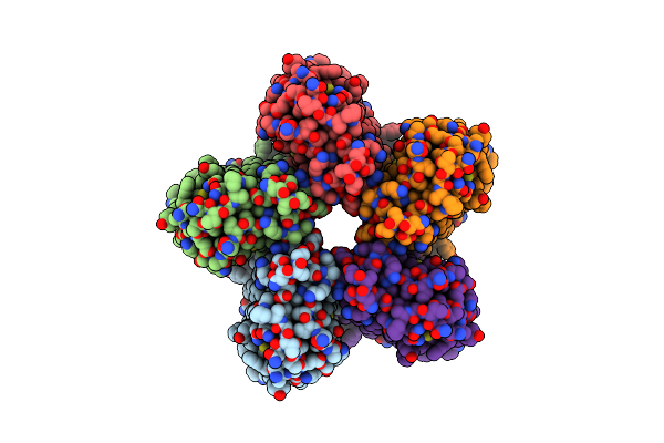
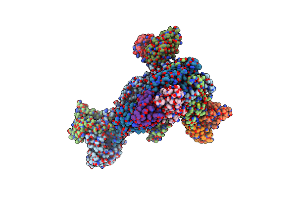
Trimeric Hsv-1F Gb Ectodomain In Postfusion Conformation With Three Bound Hdit101 Fab Molecules.
Organism: Human alphaherpesvirus 1 strain f, Homo sapiens
Method: ELECTRON MICROSCOPY Resolution:3.27 Å Release Date: 2024-06-19 Classification: VIRAL PROTEIN |
 |
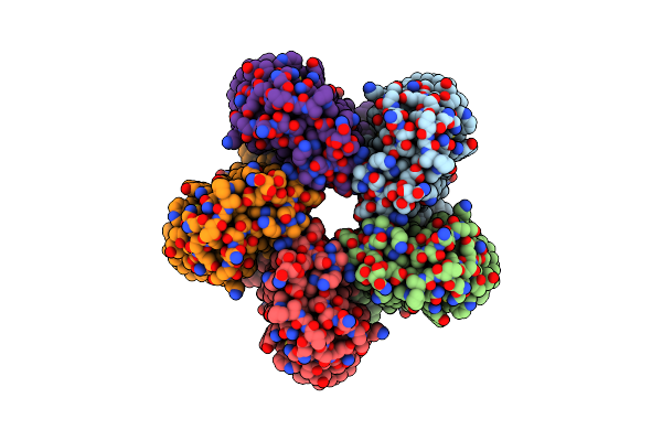
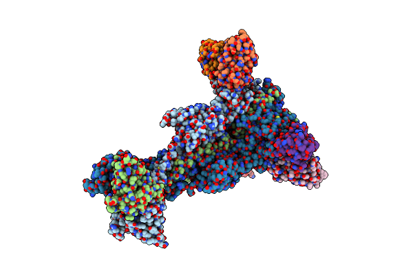
Trimeric Hsv-1F Gb Ectodomain In Postfusion Conformation With Three Bound Hdit102 Fab Molecules.
Organism: Human alphaherpesvirus 1, Homo sapiens
Method: ELECTRON MICROSCOPY Resolution:3.44 Å Release Date: 2024-06-19 Classification: VIRAL PROTEIN |
 |
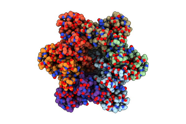
Trimeric Hsv-2F Gb Ectodomain In Postfusion Conformation With Three Bound Hdit101 Fab Molecules.
Organism: Human herpesvirus 2 strain g, Homo sapiens
Method: ELECTRON MICROSCOPY Resolution:3.45 Å Release Date: 2024-06-19 Classification: VIRAL PROTEIN |
 |
Trimeric Hsv-2G Gb Ectodomain In Postfusion Conformation With Three Bound Hdit102 Fab Molecules.
Organism: Human herpesvirus 2 strain g, Homo sapiens
Method: ELECTRON MICROSCOPY Resolution:3.12 Å Release Date: 2024-06-19 Classification: VIRAL PROTEIN |
 |
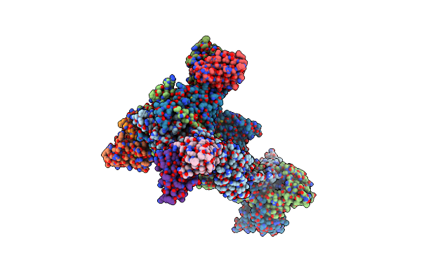
Cryo-Em Structure Of The Canine Distemper Virus Tetrameric Attachment Glycoprotein
Organism: Canine morbillivirus
Method: ELECTRON MICROSCOPY Release Date: 2023-02-08 Classification: VIRAL PROTEIN |
 |
Crystal Structure Of A Mfs Transporter With Bound 1-Hydroxynaphthalene-2-Carboxylic Acid At 2.67 Angstroem Resolution
Organism: Syntrophobacter fumaroxidans mpob
Method: X-RAY DIFFRACTION Resolution:2.46 Å Release Date: 2021-10-20 Classification: MEMBRANE PROTEIN Ligands: 1HN |
 |
Crystal Structure Of A Mfs Transporter With Bound 3-Phenylpropanoic Acid At 2.39 Angstroem Resolution
Organism: Syntrophobacter fumaroxidans mpob
Method: X-RAY DIFFRACTION Resolution:2.15 Å Release Date: 2021-10-20 Classification: MEMBRANE PROTEIN Ligands: HCI |
 |
Crystal Structure Of A Mfs Transporter With Bound 2-Naphthoic Acid At 2.39 Angstroem Resolution
Organism: Syntrophobacter fumaroxidans mpob
Method: X-RAY DIFFRACTION Resolution:2.23 Å Release Date: 2021-10-20 Classification: MEMBRANE PROTEIN Ligands: FIV |
 |
Crystal Structure Of A Mfs Transporter With Bound 3-(2-Methylphenyl)Propanoic Acid At 2.41 Angstroem Resolution
Organism: Syntrophobacter fumaroxidans mpob
Method: X-RAY DIFFRACTION Resolution:2.18 Å Release Date: 2021-10-20 Classification: MEMBRANE PROTEIN Ligands: 02Q |
 |
Organism: Escherichia coli o157:h7
Method: X-RAY DIFFRACTION Resolution:1.69 Å Release Date: 2021-09-08 Classification: TRANSPORT PROTEIN Ligands: BNG, TRS, HEX, D10, OCT |
 |
Organism: Uncultured gammaproteobacteria bacterium
Method: ELECTRON MICROSCOPY Release Date: 2021-06-16 Classification: PROTON TRANSPORT Ligands: RET |
 |
Cryo-Em Structure Of The Prefusion State Of Canine Distemper Virus Fusion Protein Ectodomain
Organism: Canine morbillivirus
Method: ELECTRON MICROSCOPY Release Date: 2020-07-22 Classification: VIRAL PROTEIN |
 |
Organism: Syntrophobacter fumaroxidans (strain dsm 10017 / mpob)
Method: X-RAY DIFFRACTION Resolution:2.30 Å Release Date: 2019-07-03 Classification: MEMBRANE PROTEIN Ligands: HG, JKE, EPE, BNG |
 |
Crystal Structure Of A Mfs Transporter With Ligand At 2.69 Angstroem Resolution
Organism: Syntrophobacter fumaroxidans mpob
Method: X-RAY DIFFRACTION Resolution:2.50 Å Release Date: 2019-07-03 Classification: MEMBRANE PROTEIN Ligands: 2OP, BNG |