
Search Count: 122
 |
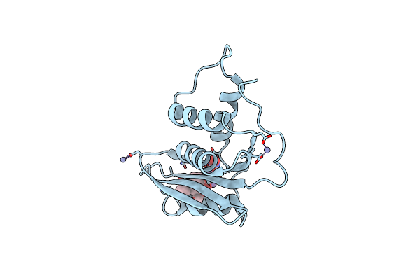
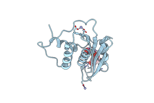
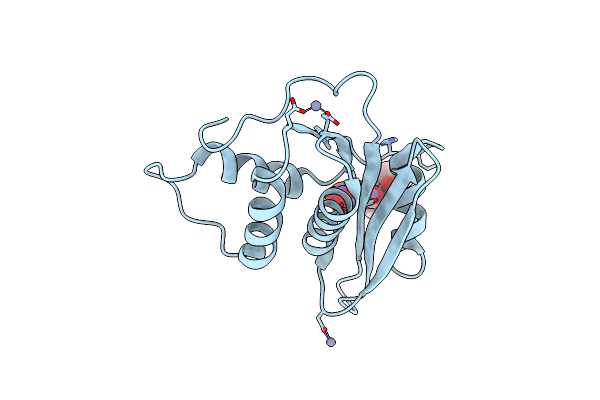
Organism: Homo sapiens
Method: X-RAY DIFFRACTION Resolution:2.50 Å Release Date: 2025-01-01 Classification: IMMUNE SYSTEM Ligands: NAG, SO4 |
 |
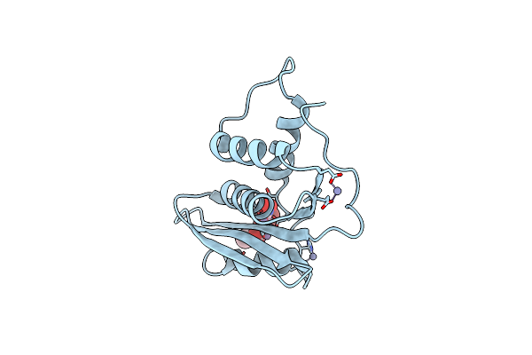
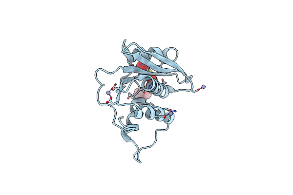
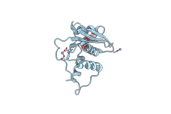
Organism: Homo sapiens
Method: X-RAY DIFFRACTION Resolution:2.60 Å Release Date: 2025-01-01 Classification: IMMUNE SYSTEM Ligands: NAG, SO4 |
 |
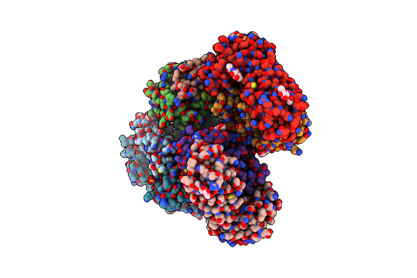
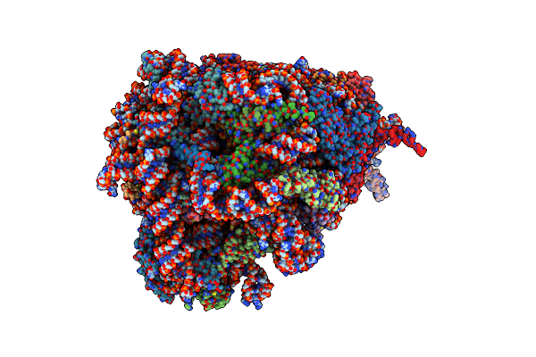
Organism: Homo sapiens
Method: ELECTRON MICROSCOPY Release Date: 2025-01-01 Classification: IMMUNE SYSTEM Ligands: 6UL, NAG, GLC, A1L2B |
 |
Organism: Homo sapiens
Method: X-RAY DIFFRACTION Resolution:2.95 Å Release Date: 2024-10-09 Classification: DNA/DNA BINDING PROTEIN Ligands: ZN |
 |
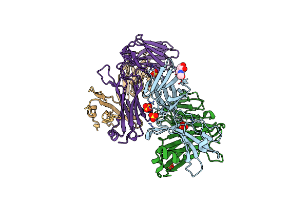
Organism: Homo sapiens, Severe acute respiratory syndrome coronavirus 2, Synthetic construct
Method: ELECTRON MICROSCOPY Resolution:3.40 Å Release Date: 2023-12-27 Classification: VIRAL PROTEIN/PROTEIN BINDING Ligands: NAG, ZN, SO4 |
 |
Organism: Homo sapiens
Method: X-RAY DIFFRACTION Resolution:1.49 Å Release Date: 2022-12-21 Classification: TRANSFERASE Ligands: ZN, SFG, J26, EDO, TRS |
 |
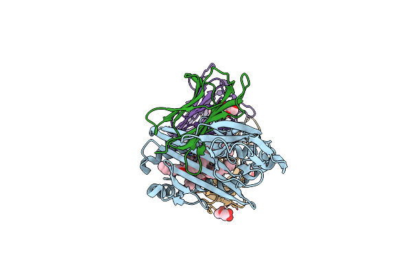
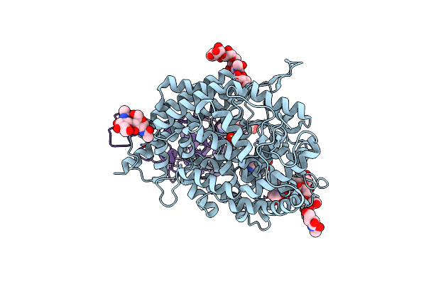
Organism: Homo sapiens
Method: ELECTRON MICROSCOPY Release Date: 2022-06-22 Classification: RIBOSOME Ligands: MG, ZN |
 |
Crystal Structure Of Engineered Hiv-1 Reverse Transcriptase Rnase H Domain Complexed With Nitrofuran Methoxy(Methoxycarbonyl)Phenyl Ester
Organism: Human immunodeficiency virus 1
Method: X-RAY DIFFRACTION Resolution:1.88 Å Release Date: 2022-04-27 Classification: VIRAL PROTEIN Ligands: MN, E58, ZN |
 |
Crystal Structure Of Engineered Hiv-1 Reverse Transcriptase Rnase H Domain Complexed With Nitrofuran Methoxy(Methoxycarbonyl)Phenyl Ester
Organism: Human immunodeficiency virus 1
Method: X-RAY DIFFRACTION Resolution:2.18 Å Release Date: 2022-04-27 Classification: VIRAL PROTEIN Ligands: MN, ZN, E6I |
 |
Crystal Structure Of Engineered Hiv-1 Reverse Transcriptase Rnase H Domain Complexed With Nitrofuran Methoxy(Methoxycarbonyl)Phenyl Ester
Organism: Human immunodeficiency virus 1
Method: X-RAY DIFFRACTION Resolution:2.09 Å Release Date: 2022-04-27 Classification: VIRAL PROTEIN Ligands: MN, E81, ZN |
 |
Crystal Structure Of Engineered Hiv-1 Reverse Transcriptase Rnase H Domain Complexed With Nitrofuran Methoxy(Methoxycarbonyl)Phenyl Ester
Organism: Human immunodeficiency virus 1
Method: X-RAY DIFFRACTION Resolution:1.80 Å Release Date: 2022-04-27 Classification: VIRAL PROTEIN Ligands: MN, ZN, E9B |
 |
Crystal Structure Of Engineered Hiv-1 Reverse Transcriptase Rnase H Domain Complexed With Nitrofuran Methoxy(Methoxycarbonyl)Phenyl Ester
Organism: Human immunodeficiency virus 1
Method: X-RAY DIFFRACTION Resolution:1.75 Å Release Date: 2022-04-27 Classification: VIRAL PROTEIN Ligands: ECW, MN, ZN |
 |
Crystal Structure Of Engineered Hiv-1 Reverse Transcriptase Rnase H Domain Complexed With Nitrofuran Methoxy(Methoxycarbonyl)Phenyl Ester
Organism: Human immunodeficiency virus 1
Method: X-RAY DIFFRACTION Resolution:1.80 Å Release Date: 2022-04-27 Classification: VIRAL PROTEIN Ligands: MN, ZN, EGI |
 |
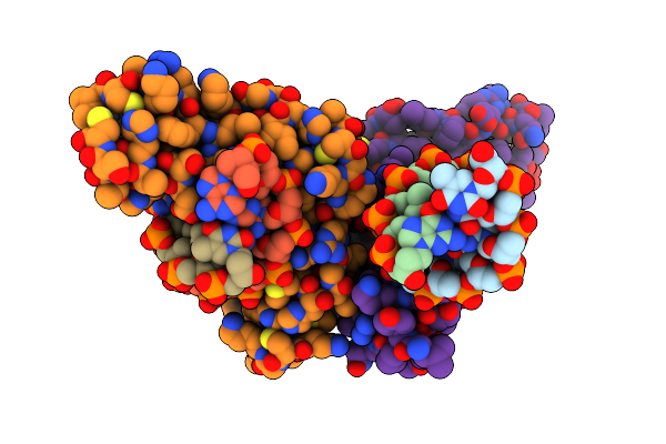
Structure Of Sars-Cov-2 Spike Receptor-Binding Domain Complexed With High Affinity Ace2 Mutant 3N39
Organism: Homo sapiens, Severe acute respiratory syndrome coronavirus 2
Method: X-RAY DIFFRACTION Resolution:3.20 Å Release Date: 2020-12-23 Classification: VIRAL PROTEIN Ligands: NAG, ZN, SO4 |
 |
Organism: Streptococcus sp. 'group g'
Method: X-RAY DIFFRACTION Resolution:1.84 Å Release Date: 2019-10-23 Classification: IMMUNE SYSTEM Ligands: ACY |
 |
Crystal Structure Of Tandem Tudor Domain Of Human Uhrf1 In Complex With Lig1-K126Me3
Organism: Homo sapiens, Synthetic construct
Method: X-RAY DIFFRACTION Resolution:2.65 Å Release Date: 2018-12-12 Classification: TRANSFERASE |
 |
Organism: Homo sapiens
Method: X-RAY DIFFRACTION Resolution:1.70 Å Release Date: 2018-12-12 Classification: TRANSFERASE Ligands: EDO, SO4 |
 |
Organism: Escherichia coli
Method: X-RAY DIFFRACTION Resolution:1.33 Å Release Date: 2016-12-14 Classification: SIGNALING PROTEIN |
 |
Organism: Escherichia coli
Method: X-RAY DIFFRACTION Resolution:2.30 Å Release Date: 2016-12-14 Classification: SIGNALING PROTEIN |
 |
Crystal Structure Of Formyltetrahydrofolate Deformylase From Thermus Thermophilus Hb8
Organism: Thermus thermophilus
Method: X-RAY DIFFRACTION Resolution:2.71 Å Release Date: 2014-01-08 Classification: HYDROLASE |