
Search Count: 2,008
 |
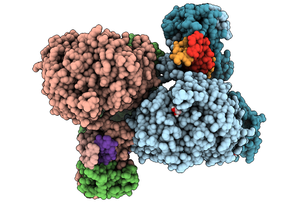
Organism: Pyrococcus furiosus
Method: ELECTRON MICROSCOPY Release Date: 2025-11-05 Classification: RIBOSOME Ligands: ZN |
 |
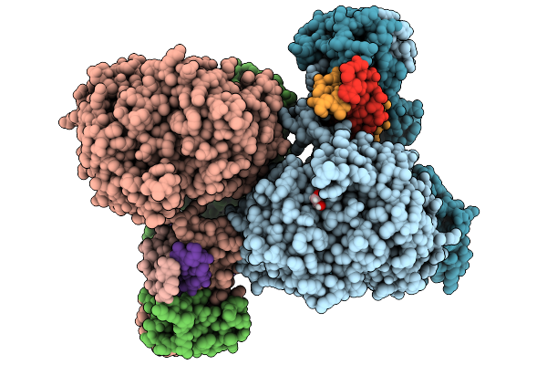
Organism: Pyrococcus furiosus
Method: ELECTRON MICROSCOPY Release Date: 2025-11-05 Classification: RIBOSOME Ligands: ZN |
 |
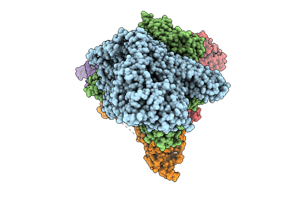
Organism: Pyrococcus furiosus
Method: ELECTRON MICROSCOPY Release Date: 2025-11-05 Classification: RIBOSOME Ligands: ZN |
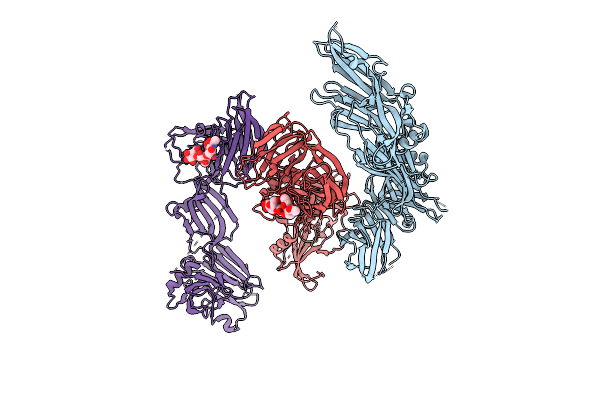 |
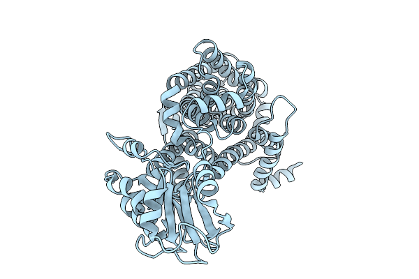
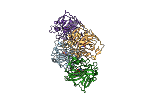
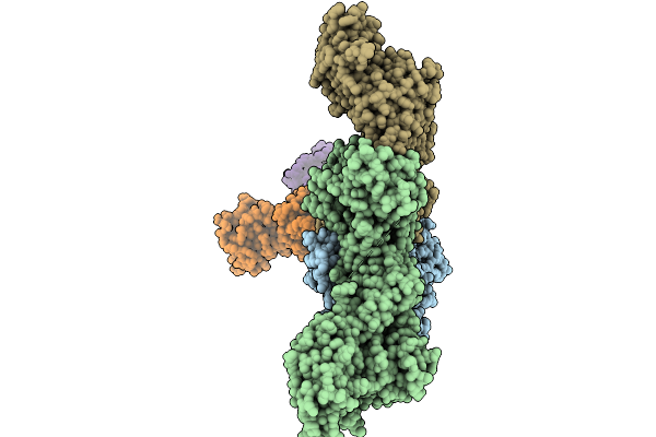
Crystal Structure Of Ha3 From Clostridium Botulinum Type B With Alpha2,3-Sialyllactose
Organism: Clostridium botulinum b1 str. okra
Method: X-RAY DIFFRACTION Release Date: 2025-11-05 Classification: TOXIN |
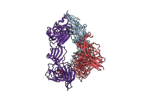 |
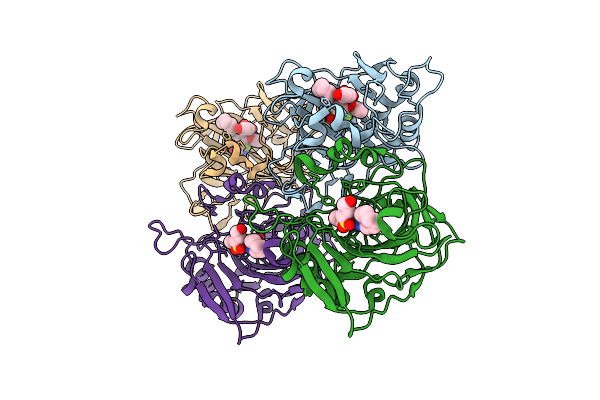
Crystal Structure Of Ha3 From Clostridium Botulinum Type B With Alpha2,6-Sialyllactose
Organism: Clostridium botulinum b1 str. okra
Method: X-RAY DIFFRACTION Release Date: 2025-11-05 Classification: TOXIN Ligands: SIA |
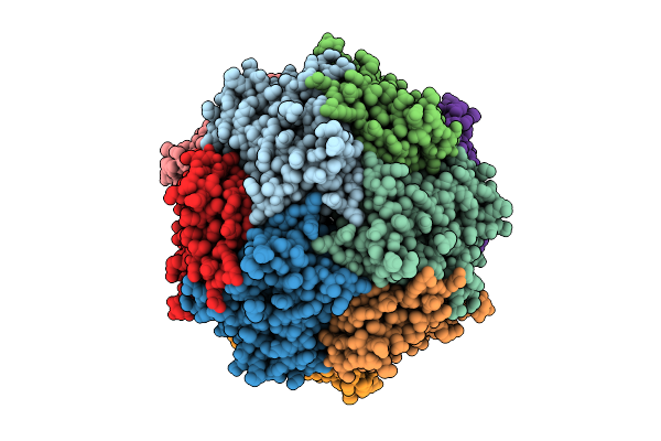 |
Structure Of Thioferritin (Pfdpsl) With Ferrihydrite Growth At A Single Three-Fold Pore.
Organism: Pyrococcus furiosus
Method: ELECTRON MICROSCOPY Release Date: 2025-10-29 Classification: METAL BINDING PROTEIN Ligands: FE, OXY, O |
 |
Organism: Homo sapiens
Method: X-RAY DIFFRACTION Release Date: 2025-10-22 Classification: ONCOPROTEIN Ligands: ZN, A1IWG |
 |
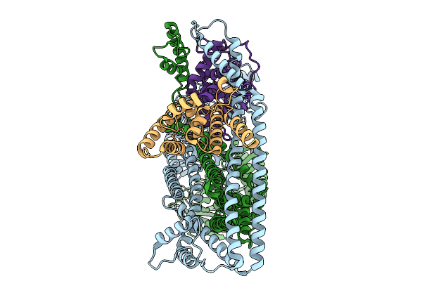
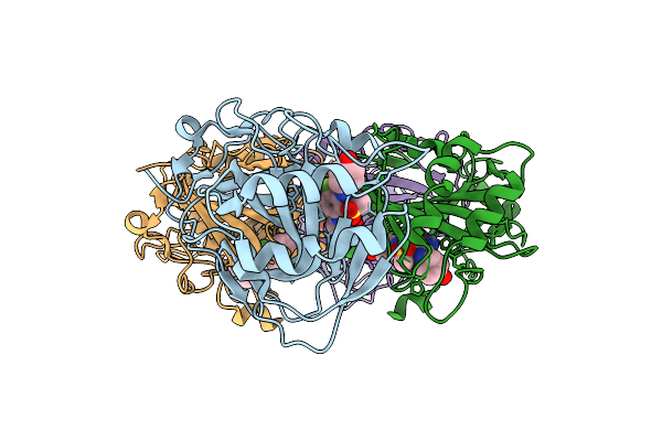
X-Ray Crystal Structure Of Asp/Ala Exchanger Aspt At Outward-Facing Conformation
Organism: Tetragenococcus halophilus
Method: X-RAY DIFFRACTION Release Date: 2025-10-08 Classification: MEMBRANE PROTEIN |
 |
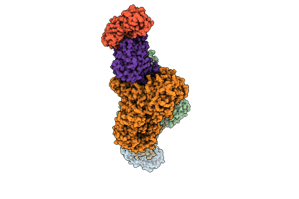
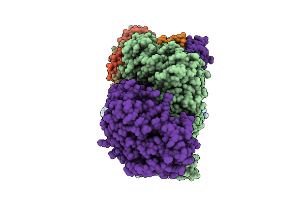
Organism: Salmonella enterica subsp. enterica serovar tennessee
Method: ELECTRON MICROSCOPY Release Date: 2025-10-01 Classification: IMMUNE SYSTEM |
 |
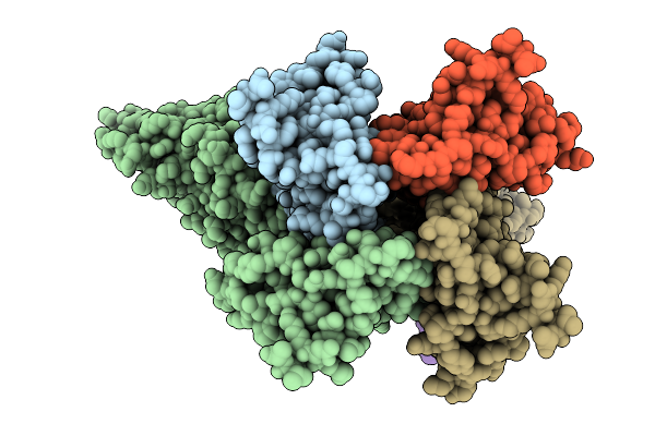
Organism: Salmonella enterica subsp. enterica serovar tennessee, Bacillus subtilis subsp. subtilis str. 168
Method: ELECTRON MICROSCOPY Release Date: 2025-10-01 Classification: IMMUNE SYSTEM |
 |
Organism: Salmonella enterica subsp. enterica serovar tennessee, Bacillus subtilis subsp. subtilis str. 168
Method: ELECTRON MICROSCOPY Release Date: 2025-10-01 Classification: IMMUNE SYSTEM |
 |
Three Dimensional Structure Of Human Carbonic Anhydrase Xii In Complex With Sulfonamide
Organism: Homo sapiens
Method: X-RAY DIFFRACTION Release Date: 2025-09-17 Classification: LYASE Ligands: ZN, A1H9A |
 |
Three Dimensional Structure Of Human Carbonic Anhydrase Xii In Complex With Sulfonamide
Organism: Homo sapiens
Method: X-RAY DIFFRACTION Release Date: 2025-09-17 Classification: LYASE Ligands: ZN, A1H92 |
 |
Three Dimensional Structure Of Human Carbonic Anhydrase Ix In Complex With Sulfonamide
Organism: Homo sapiens
Method: X-RAY DIFFRACTION Release Date: 2025-09-17 Classification: LYASE Ligands: ZN, A1JC2 |
 |
Three Dimensional Structure Of Human Carbonic Anhydrase Xii In Complex With Sulfonamide
Organism: Homo sapiens
Method: X-RAY DIFFRACTION Release Date: 2025-09-17 Classification: LYASE Ligands: ZN, A1H89 |
 |
Crystal Structure Of Mbp-Fused Bil1/Bzr1 (21-104) In Complex With Double-Stranded Dna Contaning Cacatatgtg
Organism: Arabidopsis thaliana, Synthetic construct
Method: X-RAY DIFFRACTION Release Date: 2025-09-10 Classification: DNA BINDING PROTEIN Ligands: EDO |
 |
Crystal Structure Of Mbp-Fused Bil1/Bzr1 (21-104) In Complex With Double-Stranded Dna Contaning Cacagctgtg
Organism: Arabidopsis thaliana, Synthetic construct
Method: X-RAY DIFFRACTION Release Date: 2025-09-10 Classification: DNA BINDING PROTEIN Ligands: EDO |
 |
Organism: Clostridium botulinum
Method: ELECTRON MICROSCOPY Release Date: 2025-09-10 Classification: TOXIN |
 |
Organism: Clostridium botulinum
Method: ELECTRON MICROSCOPY Release Date: 2025-09-10 Classification: TOXIN |
 |
Organism: Clostridium botulinum
Method: ELECTRON MICROSCOPY Release Date: 2025-09-10 Classification: TOXIN |