
Search Count: 46
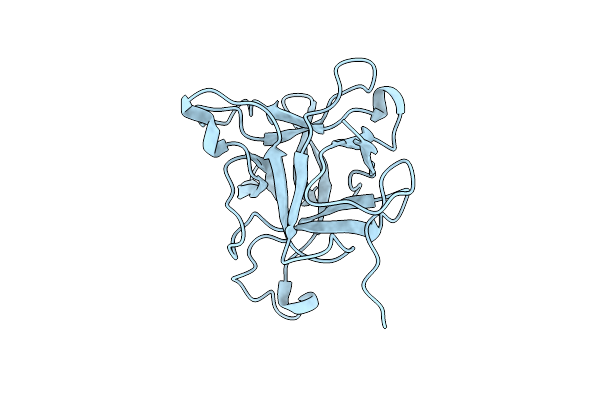 |
Crystal Structure At Ph 7.0 Of A Potato Sti-Kunitz Bi-Functional Inhibitor Of Serine And Aspartic Proteases In Space Group P4322 And Ph 9.0
Organism: Solanum tuberosum
Method: X-RAY DIFFRACTION Resolution:2.45 Å Release Date: 2016-07-06 Classification: HYDROLASE INHIBITOR |
 |
Crystal Structure At Ph 9.0 Of A Potato Sti-Kunitz Bi-Functional Inhibitor Of Serine And Aspartic Proteases In Space Group P4322 And Ph 9.0
Organism: Solanum tuberosum
Method: X-RAY DIFFRACTION Resolution:2.65 Å Release Date: 2016-07-06 Classification: HYDROLASE INHIBITOR |
 |
Crystal Structure Of N19D Potato Sti-Kunitz Bi-Functional Inhibitor Of Serine And Aspartic Proteases In Space Group C2221 And Ph 3.5
Organism: Solanum tuberosum
Method: X-RAY DIFFRACTION Resolution:2.47 Å Release Date: 2016-07-06 Classification: HYDROLASE INHIBITOR |
 |
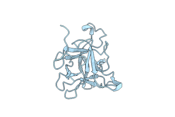
Crystal Structure Of Potato Sti-Kunitz Bi-Functional Inhibitor Of Serine And Aspartic Proteases In Space Group P22121 And Ph 7.0
Organism: Solanum tuberosum
Method: X-RAY DIFFRACTION Resolution:2.55 Å Release Date: 2016-07-06 Classification: HYDROLASE |
 |
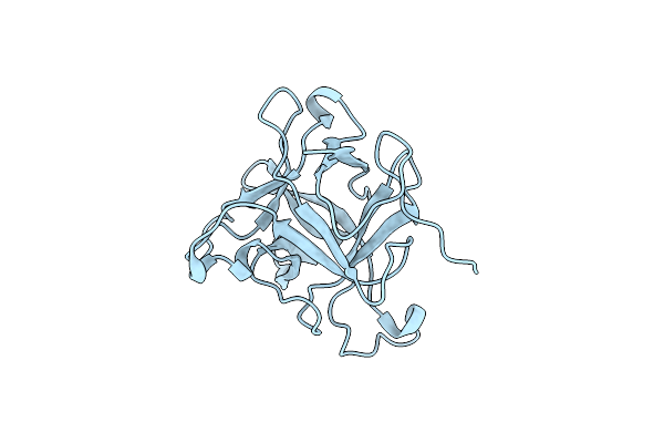
Crystal Structure Of A Potato Sti-Kunitz Bifunctional Inhibitor Of Serine And Aspartic Proteases In Space Group P4322 And Ph 7.4
Organism: Solanum tuberosum
Method: X-RAY DIFFRACTION Resolution:2.50 Å Release Date: 2016-07-06 Classification: HYDROLASE INHIBITOR Ligands: NAG |
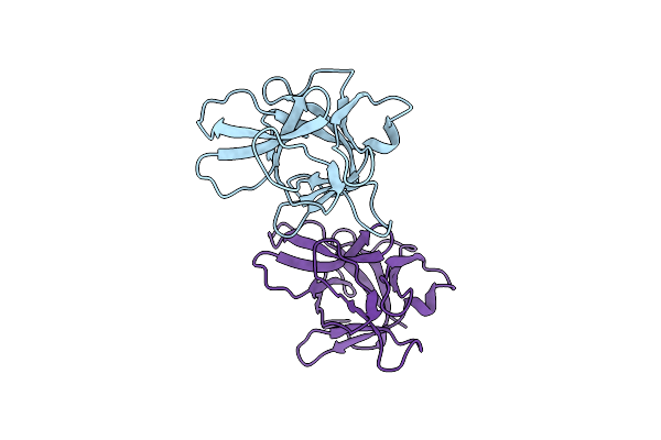 |
Organism: Clitocybe nebularis
Method: X-RAY DIFFRACTION Resolution:1.95 Å Release Date: 2009-10-20 Classification: HYDROLASE/HYDROLASE INHIBITOR |
 |
Organism: Homo sapiens, Clitocybe nebularis
Method: X-RAY DIFFRACTION Resolution:2.22 Å Release Date: 2009-10-20 Classification: HYDROLASE/HYDROLASE INHIBITOR Ligands: SO4 |
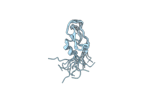 |
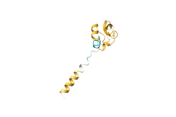
Solution Structure Of Bungarus Faciatus Ix, A Kunitz-Type Chymotrypsin Inhibitor
|
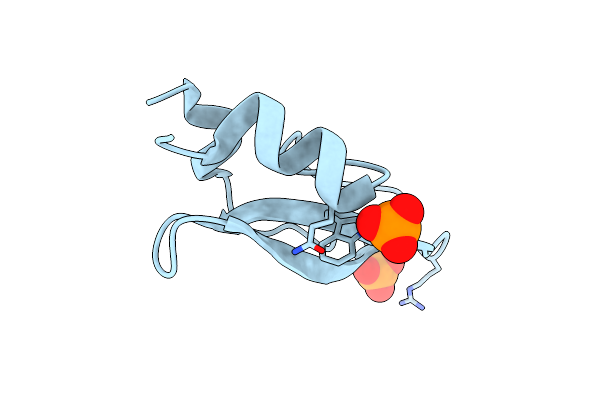 |
Organism: Homo sapiens
Method: X-RAY DIFFRACTION Resolution:0.95 Å Release Date: 2002-02-06 Classification: STRUCTURAL PROTEIN Ligands: PO4 |
 |
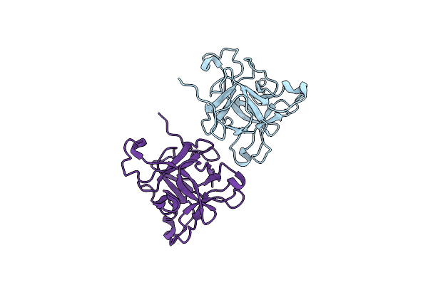
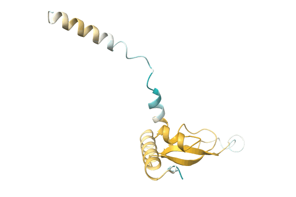
Crystal Structure Of Tick Anticoagulant Protein Complexed With Bovine Pancreatic Trypsin Inhibitor
Organism: Ornithodoros moubata, Bos taurus
Method: X-RAY DIFFRACTION Resolution:1.62 Å Release Date: 2000-09-09 Classification: BLOOD CLOTTING INHIBITOR Ligands: SO4 |
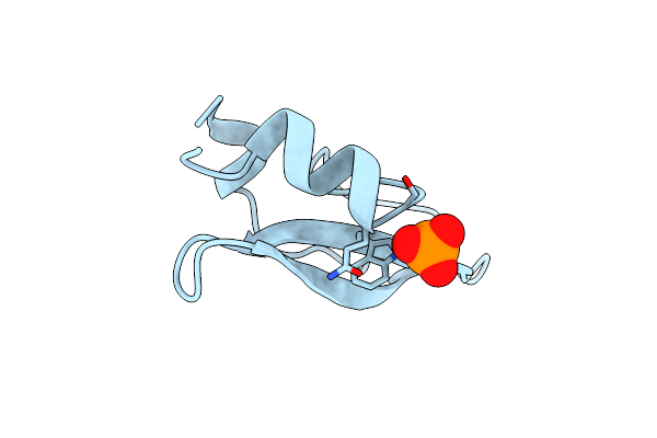 |
Organism: Homo sapiens
Method: X-RAY DIFFRACTION Resolution:1.20 Å Release Date: 1997-05-15 Classification: KUNITZ INHIBITOR Ligands: PO4 |
 |
 |
Organism: Operophtera brumata (winter moth)
Method: Alphafold Release Date: Classification: NA Ligands: NA |
 |
 |
 |
 |
 |
 |
 |