
Search Count: 24
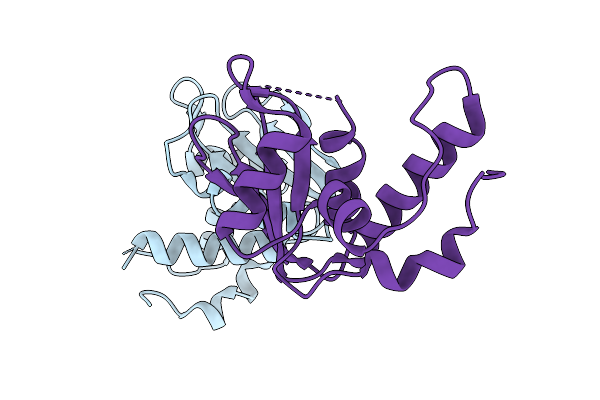 |
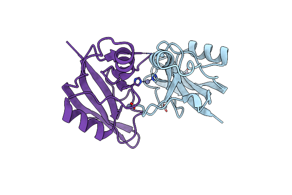
Pantoea Ananatis Encodes An Antibacterial And Anti-Eukaryotic Human Cd38 Homologue T6Ss Adp-Ribosyl Cyclase Polymorphic Toxin
Organism: Pantoea ananatis lmg 20103
Method: X-RAY DIFFRACTION Resolution:1.59 Å Release Date: 2025-10-01 Classification: TOXIN |
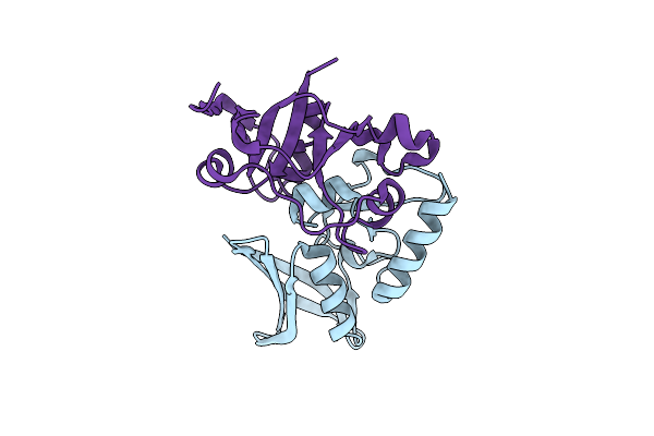 |
Xenorhabdus Bovienii Rhs C-Terminal Toxin Trex Complex With Trix Immunity Protein
Organism: Xenorhabdus bovienii ss-2004
Method: X-RAY DIFFRACTION Resolution:1.28 Å Release Date: 2024-12-04 Classification: TOXIN |
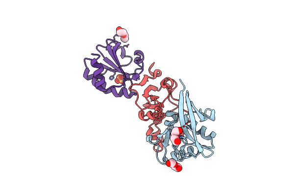 |
Xenorhabdus Bovienii Rhs Toxin Tretu Complex With Trxa And Tritu Immunity Protein
Organism: Xenorhabdus bovienii ss-2004, Escherichia coli str. k-12 substr. mg1655
Method: X-RAY DIFFRACTION Resolution:2.11 Å Release Date: 2024-12-04 Classification: TOXIN Ligands: PEG, SO4 |
 |
Xenorhabdus Bovienii Thioredoxin Complex With Rhs Toxin Trex And Immunity Protein Trix
Organism: Xenorhabdus bovienii ss-2004
Method: X-RAY DIFFRACTION Resolution:2.00 Å Release Date: 2024-12-04 Classification: TOXIN Ligands: CA, PEG |
 |
Organism: Acinetobacter baumannii
Method: X-RAY DIFFRACTION Resolution:1.85 Å Release Date: 2023-07-12 Classification: TRANSFERASE Ligands: COA, MG, LFU, GOL, 13D |
 |
Organism: Acinetobacter baumannii
Method: X-RAY DIFFRACTION Resolution:1.59 Å Release Date: 2023-07-12 Classification: TRANSFERASE Ligands: COA, BR, NA, MG, ACO |
 |
Organism: Salmonella enterica subsp. enterica serovar typhimurium
Method: X-RAY DIFFRACTION Resolution:2.20 Å Release Date: 2022-11-23 Classification: TOXIN Ligands: ZN |
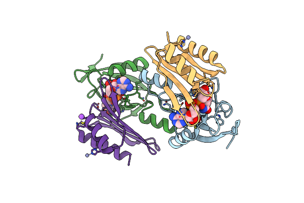 |
Salmonella Enterica Rhs1 C-Terminal Toxin Tretu Complex With Tritu Immunity Protein
Organism: Salmonella enterica subsp. enterica serovar typhimurium
Method: X-RAY DIFFRACTION Resolution:2.70 Å Release Date: 2022-11-23 Classification: TOXIN Ligands: NAD, NA, ZN |
 |
Photorhabdus Laumondii T6Ss-Associated Rhs Protein Carrying The Tre23 Toxin Domain
Organism: Photorhabdus laumondii subsp. laumondii tto1
Method: ELECTRON MICROSCOPY Resolution:3.17 Å Release Date: 2021-12-01 Classification: TOXIN |
 |
Organism: Escherichia coli str. k-12 substr. mg1655, Escherichia coli 2-222-05_s4_c3
Method: X-RAY DIFFRACTION Resolution:2.28 Å Release Date: 2019-09-18 Classification: TOXIN Ligands: SO4 |
 |
Organism: Escherichia coli 2-210-07_s3_c3
Method: X-RAY DIFFRACTION Resolution:2.26 Å Release Date: 2019-09-18 Classification: ANTITOXIN |
 |
Organism: Escherichia coli
Method: X-RAY DIFFRACTION Resolution:2.97 Å Release Date: 2019-03-06 Classification: TRANSCRIPTION |
 |
Organism: Escherichia coli
Method: X-RAY DIFFRACTION Resolution:2.50 Å Release Date: 2019-03-06 Classification: TRANSCRIPTION Ligands: ACO, MG, CL |
 |
Structure Of The Atat Y144F Mutant Toxin Bound To The C-Terminus Of The Antitoxin Atar
Organism: Escherichia coli
Method: X-RAY DIFFRACTION Resolution:2.49 Å Release Date: 2019-03-06 Classification: TRANSCRIPTION Ligands: CL, GOL, PO4, ACT, FLC, IOD |
 |
Structure Of The Atat Y144F Mutant Toxin Bound To The C-Terminus Of The Antitoxin Atar And Acetyl-Coa
Organism: Escherichia coli
Method: X-RAY DIFFRACTION Resolution:2.99 Å Release Date: 2019-03-06 Classification: TRANSCRIPTION Ligands: ACO, MG, GOL |
 |
Organism: Escherichia coli
Method: X-RAY DIFFRACTION Resolution:3.36 Å Release Date: 2019-03-06 Classification: TRANSCRIPTION |
 |
Structure Of Phosphorylated Translation Elongation Factor Ef-Tu From E. Coli
Organism: Escherichia coli (strain k12)
Method: X-RAY DIFFRACTION Resolution:2.80 Å Release Date: 2017-12-20 Classification: HYDROLASE Ligands: MG, GDP |
 |
Organism: Escherichia coli hs
Method: X-RAY DIFFRACTION Resolution:2.18 Å Release Date: 2017-12-20 Classification: HYDROLASE Ligands: MG, GDP, ACT, EPE, BME, CL |
 |
Organism: Escherichia coli o9:h4 (strain hs)
Method: X-RAY DIFFRACTION Resolution:3.30 Å Release Date: 2017-12-20 Classification: HYDROLASE Ligands: MG, GDP |
 |
Organism: Vibrio cholerae
Method: X-RAY DIFFRACTION Resolution:1.80 Å Release Date: 2017-04-05 Classification: ANTITOXIN |