
Search Count: 38
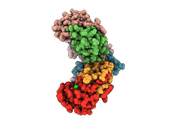 |
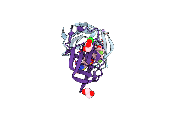
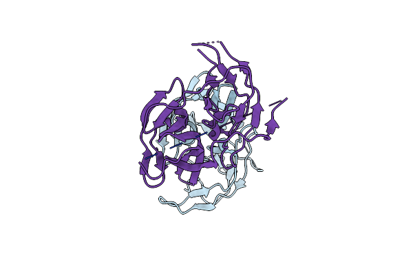
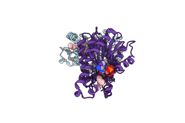
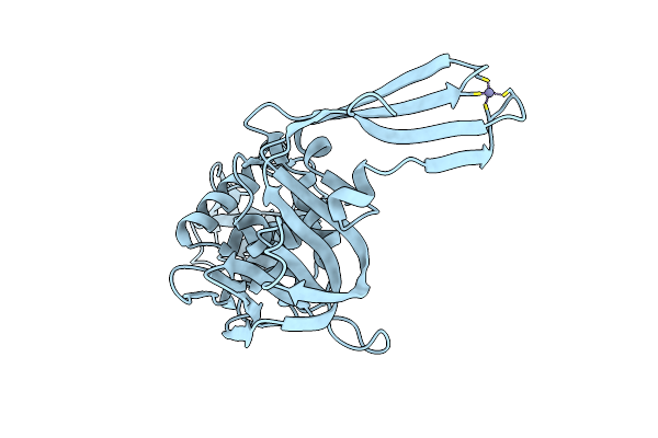
Room Temperature X-Ray Structure Of Hiv-1 Protease In Complex With Inhibitor Grl-075-24A
Organism: Human immunodeficiency virus 1
Method: X-RAY DIFFRACTION Release Date: 2025-07-23 Classification: HYDROLASE/HYDROLASE INHIBITOR Ligands: A1CAJ, CL |
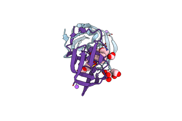 |
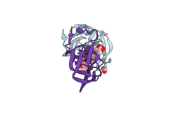
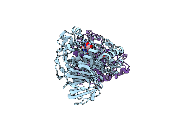
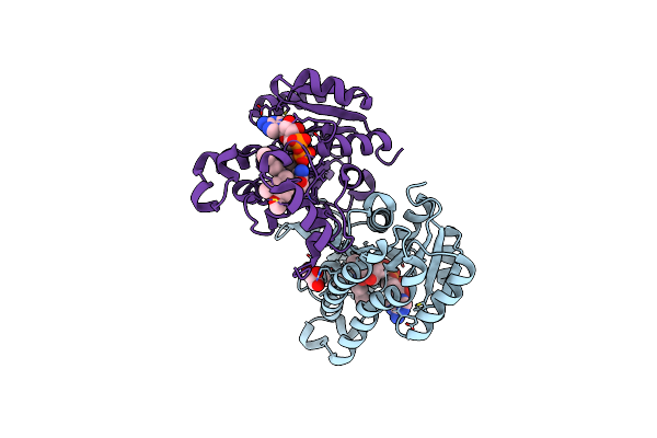
Hiv-1 Wild Type Protease With Grl-072-17A, A Substituted Tetrahydrofuran Derivative Based On Darunavir As P2 Group
Organism: Human immunodeficiency virus 1
Method: X-RAY DIFFRACTION Resolution:1.32 Å Release Date: 2024-07-24 Classification: VIRAL PROTEIN |
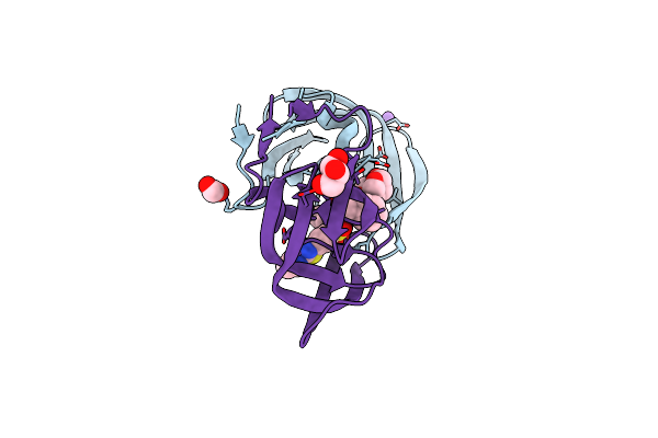 |
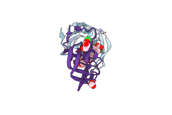
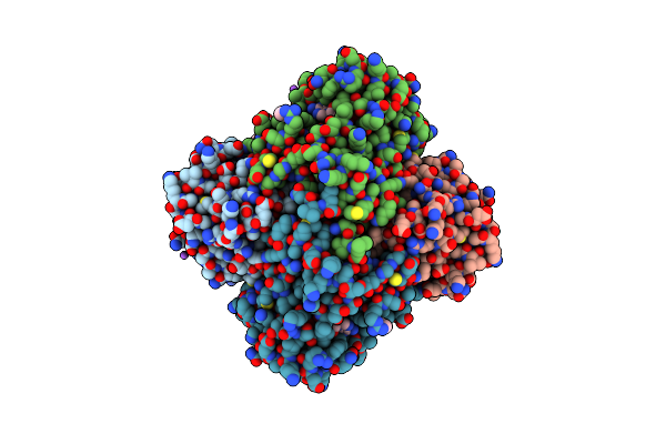
Hiv-1 Wild Type Protease With Grl-05816A, With C-4 Substituted Cyclohexane-Fused Bis-Tetrahydrofuran (Chf-Thf) Derivatives As P2-Ligand [Diastereomer 1]
Organism: Human immunodeficiency virus 1
Method: X-RAY DIFFRACTION Resolution:1.13 Å Release Date: 2022-03-02 Classification: ANTIVIRAL PROTEIN/INHIBITOR |
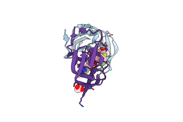 |
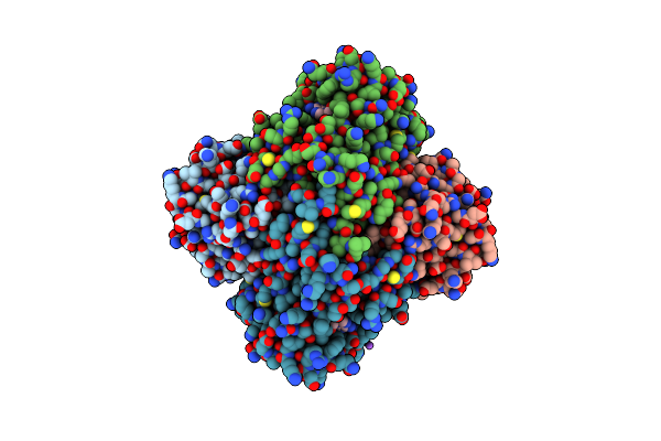
Hiv-1 Wild Type Protease With Grl-01717A, With C-4 Substituted Cyclohexane-Fused Bis-Tetrahydrofuran (Chf-Thf) Derivatives As P2-Ligand [Diastereomer 2]
Organism: Human immunodeficiency virus 1
Method: X-RAY DIFFRACTION Resolution:1.21 Å Release Date: 2022-03-02 Classification: VIRAL PROTEIN/INHIBITOR |
 |
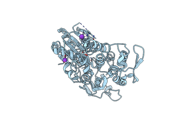
Hiv-1 Wild Type Protease With Grl-026-18A, A Crown-Like Tetrahydropyranotetrahydrofuran With A Bridged Methylene Group As A P2 Ligand
Organism: Human immunodeficiency virus 1
Method: X-RAY DIFFRACTION Resolution:1.18 Å Release Date: 2020-07-01 Classification: HYDROLASE/HYDROLASE INHIBITOR |
 |
Hiv-1 Wild Type Protease With Grl-00819A, With Phenyl-Boronic-Acid As P2'-Ligand And With A 6-5-5-Ring Fused Crown-Like Tetrahydropyranofuran As The P2-Ligand
Organism: Human immunodeficiency virus 1
Method: X-RAY DIFFRACTION Resolution:1.33 Å Release Date: 2019-10-09 Classification: VIRAL PROTEIN |
 |
Hiv-1 Wild Type Protease With Grl-03119A, With Phenyl-Boronic-Acid As P2'-Ligand And With A Hexahydro-4H-Furo-Pyran As The P2-Ligand
Organism: Human immunodeficiency virus 1
Method: X-RAY DIFFRACTION Resolution:1.13 Å Release Date: 2019-10-09 Classification: VIRAL PROTEIN Ligands: B4R, NA, CL, FMT, GOL |
 |
Organism: Zika virus (strain mr 766)
Method: X-RAY DIFFRACTION Resolution:2.05 Å Release Date: 2016-12-14 Classification: HYDROLASE Ligands: K, TRS |
 |
 |
Organism: Bacillus anthracis
Method: X-RAY DIFFRACTION Resolution:2.45 Å Release Date: 2016-08-10 Classification: HYDROLASE Ligands: ZN, NCD |
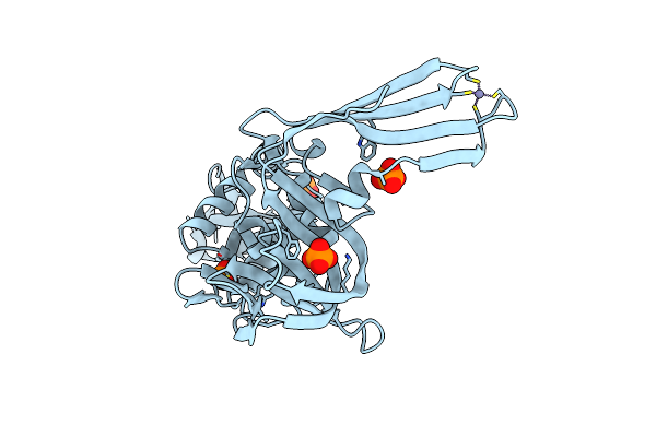 |
Crystal Structure Of Plpro From Middle East Respiratory Syndrome (Mers) Coronavirus
Organism: Human betacoronavirus 2c emc/2012
Method: X-RAY DIFFRACTION Resolution:1.79 Å Release Date: 2015-03-25 Classification: HYDROLASE Ligands: ZN, PO4 |
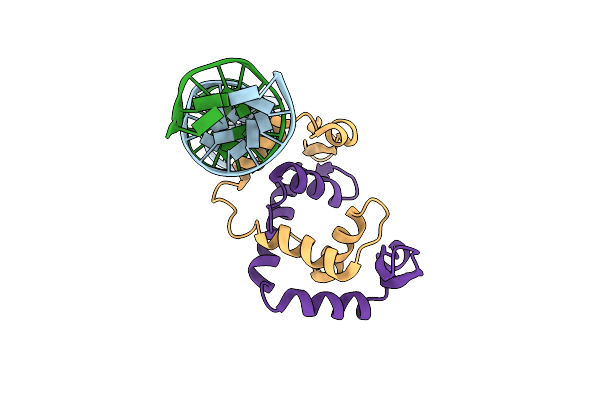 |
Organism: Homo sapiens, Synthetic construct
Method: X-RAY DIFFRACTION Resolution:3.40 Å Release Date: 2015-01-21 Classification: TRANSCRIPTION/DNA |
 |
Crystal Structure Of Fabi From S. Aureus In Complex With A Novel Benzimidazole Inhibitor
Organism: Staphylococcus aureus
Method: X-RAY DIFFRACTION Resolution:2.30 Å Release Date: 2014-12-31 Classification: oxidoreductase/oxidoreductase inhibitor Ligands: 1JT, NAP |
 |
Crystal Structures Of Fabi From F. Tularensis In Complex With Novel Inhibitors Based On The Benzimidazole Scaffold
Organism: Francisella tularensis subsp. tularensis
Method: X-RAY DIFFRACTION Resolution:2.45 Å Release Date: 2014-07-23 Classification: OXIDOREDUCTASE/OXIDOREDUCTASE INHIBITOR Ligands: NAD, 1JN, GOL |
 |
Crystal Structure Of Fabi From F. Tularensis In Complex With Novel Inhibitors Based On The Benzimidazole Scaffold.
Organism: Francisella tularensis subsp. tularensis
Method: X-RAY DIFFRACTION Resolution:1.85 Å Release Date: 2014-07-23 Classification: OXIDOREDUCTASE/OXIDOREDUCTASE INHIBITOR Ligands: 1JT, NAD, GOL, ACT, NA |
 |
Crystal Structure Of Fabi From F. Tularensis In Complex With Novel Inhibitors Based On The Benzimidazole Scaffold
Organism: Francisella tularensis subsp. tularensis
Method: X-RAY DIFFRACTION Resolution:2.34 Å Release Date: 2014-07-23 Classification: OXIDOREDUCTASE/OXIDOREDUCTASE INHIBITOR Ligands: NAD, 1JU, GOL, NA, ACT |
 |
Crystal Structure Of Plpro From Middle East Respiratory Syndrome (Mers) Coronavirus
Organism: Human betacoronavirus 2c emc/2012
Method: X-RAY DIFFRACTION Resolution:2.59 Å Release Date: 2014-05-21 Classification: HYDROLASE Ligands: ZN |
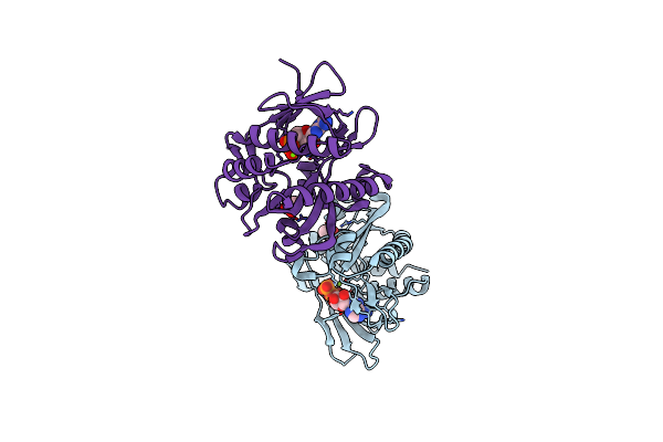 |
Structures Of Saicar Synthetase (Purc) From Streptococcus Pneumoniae With Adp, Mg2+, Air And L-Asp
Organism: Streptococcus pneumoniae
Method: X-RAY DIFFRACTION Resolution:2.69 Å Release Date: 2014-04-02 Classification: LIGASE Ligands: MG, ADP, CL, ACT |
 |
Crystal Structure Of Porphyromonas Gingivalis Enoyl-Acp Reductase Ii (Fabk) With Cofactors Nadph And Fmn
Organism: Porphyromonas gingivalis
Method: X-RAY DIFFRACTION Resolution:1.94 Å Release Date: 2014-01-29 Classification: OXIDOREDUCTASE Ligands: FMN, NDP, GOL, NA |
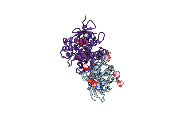 |
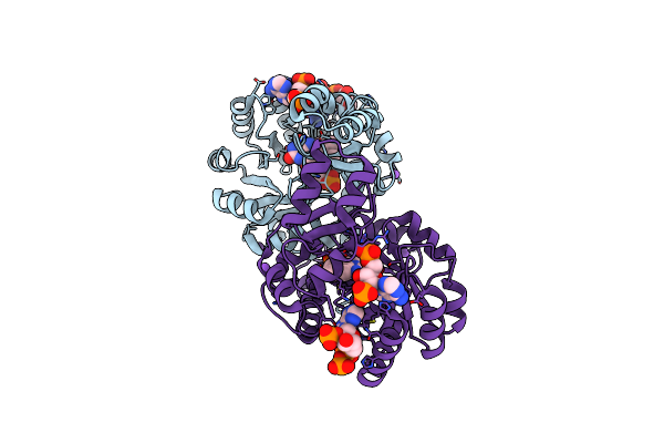
X-Ray Structure Of Saicar Synthetase (Purc) From Streptococcus Pneumoniae Complexed With Air, Adp, Asp And Mg2+
Organism: Streptococcus pneumoniae
Method: X-RAY DIFFRACTION Resolution:2.29 Å Release Date: 2013-05-29 Classification: LIGASE Ligands: AIR, ADP, MG, 144, CL, ASP, ACT |