
Search Count: 24
 |
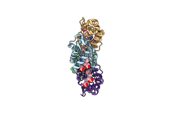
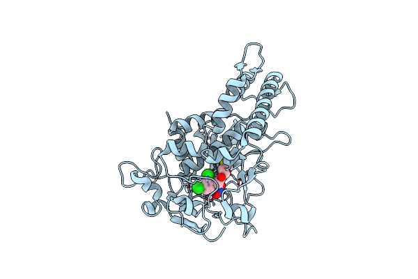
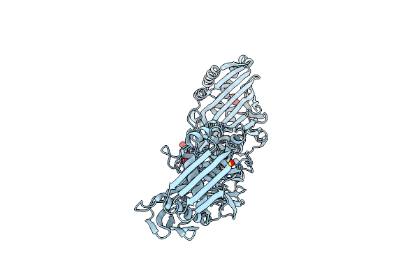
Crystal Structure Of Shewanella Benthica Group 1 Truncated Hemoglobin C51S C71S Y108A Variant
Organism: Shewanella benthica kt99
Method: X-RAY DIFFRACTION Resolution:1.70 Å Release Date: 2024-04-03 Classification: HEME BINDING PROTEIN Ligands: HEM, CYN |
 |
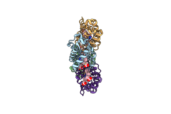
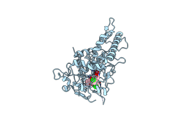
Crystal Structure Of Shewanella Benthica Group 1 Truncated Hemoglobin C51S C71S Variant
Organism: Shewanella benthica kt99
Method: X-RAY DIFFRACTION Resolution:1.80 Å Release Date: 2024-04-03 Classification: HEME-BINDING PROTEIN Ligands: HEM, CYN |
 |
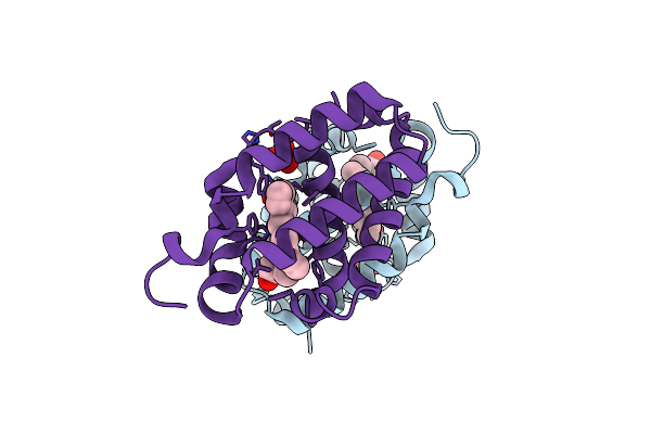
Crystal Structure Of Shewanella Benthica Group 1 Truncated Hemoglobin L80A C51S C71S Variant
Organism: Shewanella benthica kt99
Method: X-RAY DIFFRACTION Resolution:1.90 Å Release Date: 2024-04-03 Classification: HEME-BINDING PROTEIN Ligands: HEM, CYN |
 |
Crystal Structure Of Shewanella Benthica Group 1 Truncated Hemoglobin Y34F C51S C71S Variant (Cyanomet)
Organism: Shewanella benthica kt99
Method: X-RAY DIFFRACTION Resolution:1.35 Å Release Date: 2024-04-03 Classification: HEME-BINDING PROTEIN Ligands: HEM, CYN |
 |
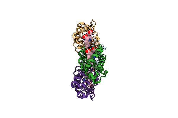
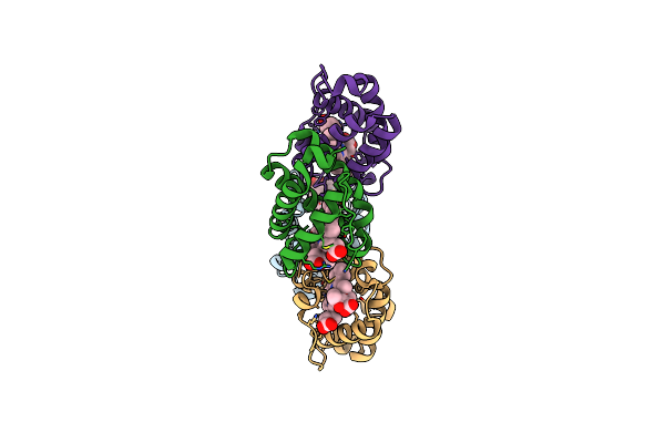
Crystal Structure Of Shewanella Benthica Group 1 Truncated Hemoglobin C51S C71S Variant With Trans Heme D
Organism: Shewanella benthica kt99
Method: X-RAY DIFFRACTION Resolution:2.00 Å Release Date: 2024-04-03 Classification: HEME-BINDING PROTEIN Ligands: A1ADT |
 |
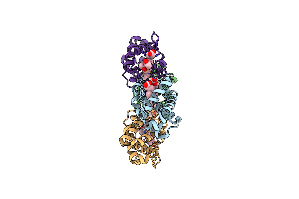
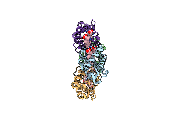
Crystal Structure Of Shewanella Benthica Group 1 Truncated Hemoglobin C51S C71S Variant With Trans Heme D
Organism: Shewanella benthica kt99
Method: X-RAY DIFFRACTION Resolution:1.80 Å Release Date: 2024-04-03 Classification: HEME-BINDING PROTEIN Ligands: HEM, A1ADT |
 |
Crystal Structure Of Shewanella Benthica Group 1 Truncated Hemoglobin C51S C71S Y34F Variant
Organism: Shewanella benthica kt99
Method: X-RAY DIFFRACTION Resolution:2.00 Å Release Date: 2022-02-16 Classification: HEME BINDING PROTEIN Ligands: HEM |
 |
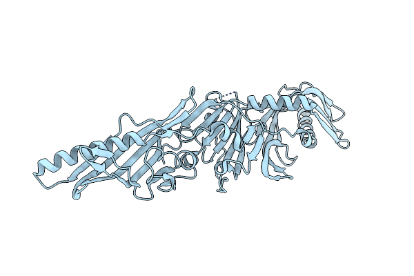
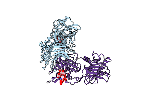
Structure Of The C. Botulinum Neurotoxin Serotype A Light Chain Protease In Complex With Covalent Inhibitor 22
Organism: Clostridium botulinum
Method: X-RAY DIFFRACTION Resolution:1.92 Å Release Date: 2021-06-09 Classification: TOXIN/INHIBITOR Ligands: ZN, UZS |
 |
Structure Of The C. Botulinum Neurotoxin Serotype A Light Chain Protease In Complex With Covalent Inhibitor 20
Organism: Clostridium botulinum
Method: X-RAY DIFFRACTION Resolution:1.74 Å Release Date: 2020-09-23 Classification: TOXIN/INHIBITOR Ligands: ZN, UZY |
 |
Structure Of The C. Botulinum Neurotoxin Serotype A Light Chain Protease In Complex With Covalent Inhibitor 21
Organism: Clostridium botulinum
Method: X-RAY DIFFRACTION Resolution:1.90 Å Release Date: 2020-09-23 Classification: TOXIN/INHIBITOR Ligands: ZN, UZV |
 |
Structure Of The C. Botulinum Neurotoxin Serotype A Light Chain Protease In Complex With Covalent Inhibitor 53
Organism: Clostridium botulinum
Method: X-RAY DIFFRACTION Resolution:2.50 Å Release Date: 2020-09-23 Classification: TOXIN/INHIBITOR Ligands: UZM |
 |
Structure Of The C. Botulinum Neurotoxin Serotype A Light Chain Protease In Complex With Noncovalent Inhibitor 59
Organism: Clostridium botulinum
Method: X-RAY DIFFRACTION Resolution:1.68 Å Release Date: 2020-09-23 Classification: TOXIN/INHIBITOR Ligands: UZP, ZN |
 |
Organism: Chlamydomonas reinhardtii
Method: X-RAY DIFFRACTION Resolution:1.90 Å Release Date: 2017-12-20 Classification: Heme Binding Protein Ligands: HEM, SO4 |
 |
Organism: Clostridium botulinum (strain kyoto / type a2)
Method: X-RAY DIFFRACTION Resolution:1.75 Å Release Date: 2017-10-25 Classification: LIPID BINDING PROTEIN |
 |
Organism: Clostridium botulinum (strain kyoto / type a2)
Method: X-RAY DIFFRACTION Resolution:2.10 Å Release Date: 2017-10-25 Classification: LIPID BINDING PROTEIN Ligands: SO4 |
 |
Organism: Clostridium botulinum
Method: X-RAY DIFFRACTION Resolution:2.59 Å Release Date: 2017-08-30 Classification: HYDROLASE Ligands: SIA |
 |
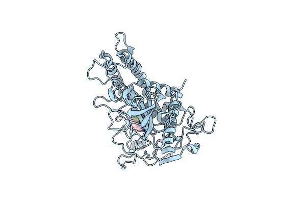
Small Molecule Inhibitor Abs-143 Bound To The Botulinum Neurotoxin Serotype A Light Chain
Organism: Clostridium botulinum
Method: X-RAY DIFFRACTION Resolution:2.50 Å Release Date: 2017-07-26 Classification: HYDROLASE/HYDROLASE INHIBITOR Ligands: ZN, 90G |
 |
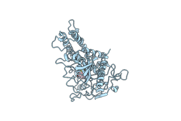
Small Molecule Inhibitor Abs-143 Bound To The Botulinum Neurotoxin Serotype A Light Chain
Organism: Clostridium botulinum
Method: X-RAY DIFFRACTION Resolution:1.90 Å Release Date: 2017-07-26 Classification: HYDROLASE/HYDROLASE INHIBITOR Ligands: ZN, 90J |
 |
Small Molecule Inhibitor Abs-143 Bound To The Botulinum Neurotoxin Serotype A Light Chain
Organism: Clostridium botulinum
Method: X-RAY DIFFRACTION Resolution:2.05 Å Release Date: 2017-07-26 Classification: HYDROLASE/HYDROLASE INHIBITOR Ligands: ZN, 90M |
 |
Organism: Clostridium botulinum
Method: X-RAY DIFFRACTION Resolution:2.60 Å Release Date: 2017-05-31 Classification: TOXIN Ligands: CU, ACT |