
Search Count: 2,756
 |
Crystal Structure Of N-Acetyl Transferase Domain-Containing Protein From Bacteroides Fragilis
Organism: Bacteroides fragilis
Method: X-RAY DIFFRACTION Release Date: 2025-09-10 Classification: TRANSFERASE Ligands: COA, SO4, EDO, ACT |
 |
Crystal Structure Of Putative Restriction Endonuclease Domain-Containing Protein From Leptospirillum Ferriphilum Ysk
Organism: Leptospirillum ferriphilum ysk
Method: X-RAY DIFFRACTION Release Date: 2025-09-10 Classification: HYDROLASE Ligands: FMT, MG, CL |
 |
Crystal Structure Of The Sars-Cov-2 2'-O-Methyltransferase With (M7Gpppa)Pupu (Cap-0) And S-Adenosyl-L-Homocysteine (Sah).
Organism: Severe acute respiratory syndrome coronavirus 2
Method: X-RAY DIFFRACTION Release Date: 2025-07-02 Classification: TRANSFERASE, Viral Protein Ligands: SAH, CL, SO4, ZN, MGT |
 |
Crystal Structure Of The Espe7 Thioesterase Mutant R35A From The Esperamicin Biosynthetic Pathway At 1.6 A
Organism: Actinomadura verrucosospora
Method: X-RAY DIFFRACTION Release Date: 2025-06-25 Classification: HYDROLASE Ligands: K, DCC |
 |
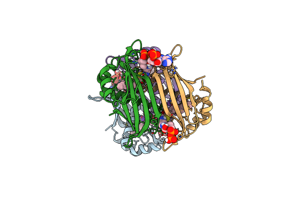
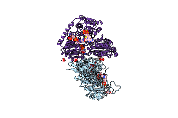
Crystal Structure Of The Complex Of M. Tuberculosis Phers With Cognate Precursor Trna And Fragment Ddd00805735
Organism: Mycobacterium tuberculosis h37rv, Mycobacterium tuberculosis (strain atcc 25618 / h37rv)
Method: X-RAY DIFFRACTION Resolution:2.51 Å Release Date: 2025-06-04 Classification: LIGASE/RNA Ligands: A1BA3, GOL, PGE, MG, NA, EPE, ACT, DMS |
 |
Crystal Structure Of The Complex Of M. Tuberculosis Phers With Cognate Precursor Trna And Fragment Ddd00107555
Organism: Mycobacterium tuberculosis h37rv
Method: X-RAY DIFFRACTION Resolution:2.05 Å Release Date: 2025-06-04 Classification: LIGASE/RNA Ligands: A1BCB, MG, ACT, NA, GOL, DMS, PGE, EPE |
 |
Crystal Structure Of The Complex Of M. Tuberculosis Phers With Cognate Precursor Trna And Fragment Ddd01008876
Organism: Mycobacterium tuberculosis h37rv
Method: X-RAY DIFFRACTION Resolution:2.45 Å Release Date: 2025-06-04 Classification: LIGASE/RNA Ligands: MG, A1BCA, ACT, NA, GOL, DMS, PGE, EPE |
 |
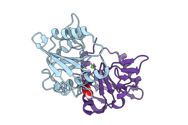
Crystal Structure Of Spermin/Spermidine N-Acetyltransferase From Enterococcus Faecalis V583
Organism: Enterococcus faecalis v583
Method: X-RAY DIFFRACTION Resolution:1.50 Å Release Date: 2025-05-28 Classification: TRANSFERASE Ligands: ACT, CL, NA, K |
 |
Organism: Staphylococcus aureus subsp. aureus usa300_fpr3757
Method: X-RAY DIFFRACTION Resolution:2.00 Å Release Date: 2025-05-14 Classification: HYDROLASE Ligands: FUM, FMT, SO4, CL |
 |
Crystal Structure Of M. Tuberculosis Phers-Trna Complex Bound To Inhibitor D-116
Organism: Mycobacterium tuberculosis h37rv
Method: X-RAY DIFFRACTION Resolution:2.35 Å Release Date: 2025-02-12 Classification: LIGASE/RNA Ligands: AX7, MG, ACT, EDO, PEG, EPE, DMS |
 |
Crystal Structure Of M. Tuberculosis Phers-Trna Complex Bound To Inhibitor D-004
Organism: Mycobacterium tuberculosis h37rv, Mycobacterium tuberculosis (strain atcc 25618 / h37rv)
Method: X-RAY DIFFRACTION Resolution:2.46 Å Release Date: 2025-02-12 Classification: LIGASE Ligands: 2AQ, MG, ACT, PEG, GOL, DMS, EPE, EDO |
 |
Crystal Structure Of Bacterial Extracellular Solute-Binding Protein From Bordetella Bronchiseptica Rb50
Organism: Bordetella bronchiseptica
Method: X-RAY DIFFRACTION Resolution:1.55 Å Release Date: 2024-12-18 Classification: STRUCTURAL GENOMICS Ligands: GOL, ACT, PGE, PG4 |
 |
Organism: Streptococcus pneumoniae
Method: X-RAY DIFFRACTION Resolution:1.62 Å Release Date: 2024-10-09 Classification: HYDROLASE Ligands: EDO, SO4 |
 |
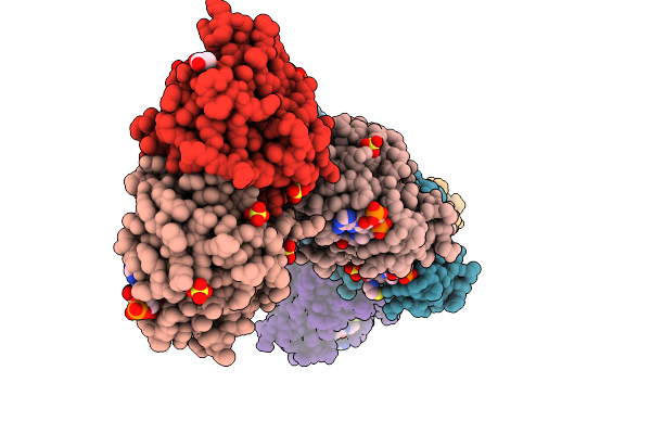
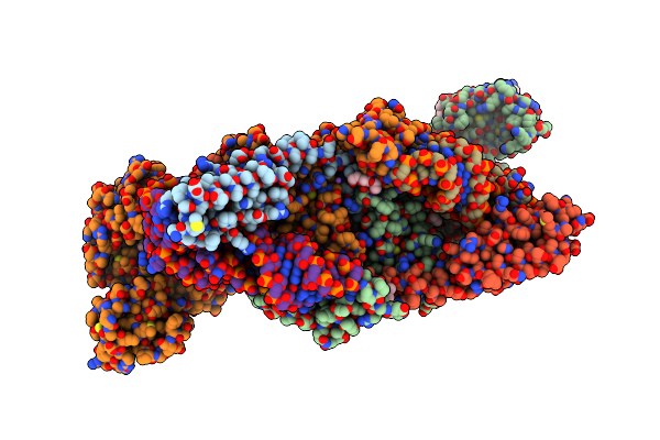
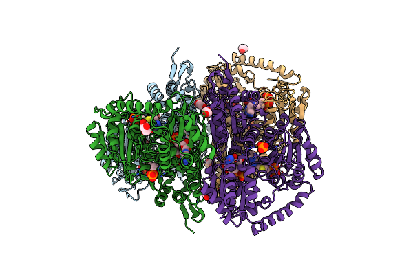
Crystal Structure Of 2-Hydroxacyl-Coa Lyase/Synthase Apbhacs From Alphaproteobacteria Bacterium In The Complex With Thdp, L-Lactyl-Coa, And Adp
Organism: Alphaproteobacteria bacterium
Method: X-RAY DIFFRACTION Resolution:2.05 Å Release Date: 2024-10-02 Classification: LYASE Ligands: 8FL, UQ3, ADP, ACE, MG, GOL |
 |
Crystal Structure Of 2-Hydroxacyl-Coa Lyase/Synthase Apbhacs From Alphaproteobacteria Bacterium In The Complex With Thdp, D-Lactyl-Coa, And Adp
Organism: Alphaproteobacteria bacterium
Method: X-RAY DIFFRACTION Resolution:1.98 Å Release Date: 2024-10-02 Classification: LYASE Ligands: MG, 8FL, UQ3, ADP, EDO, ACY, PO4, CL, ACE, PEG |
 |
Crystal Structure Of 2-Hydroxyacyl-Coa Lyase/Synthse Apbhacs From Alphaproteobacteria Bacterium In The Complex With Thdp, Formyl-Coa, And Adp
Organism: Alphaproteobacteria bacterium
Method: X-RAY DIFFRACTION Resolution:1.72 Å Release Date: 2024-10-02 Classification: LYASE Ligands: A1AEK, FYN, MG, ADP, EDO, PO4, PEG |
 |
Crystal Structure Of 2-Hydroxyacyl-Coa Lyase/Synthase Tbhacs From Thermoflexaceae Bacterium In The Complex With Thdp, Formyl-Coa And Adp
Organism: Thermoflexaceae bacterium
Method: X-RAY DIFFRACTION Resolution:2.20 Å Release Date: 2024-10-02 Classification: LYASE Ligands: TPP, FYN, CA, ADP, EDO, PEG |
 |
Crystal Structure Of 2-Hydroxyacyl-Coa Lyase/Synthase Tbhacs From Thermoflexaceae Bacterium In The Complex With Thdp, 2-Hydroxyisobutyryl-Coa And Adp
Organism: Thermoflexaceae bacterium
Method: X-RAY DIFFRACTION Resolution:1.69 Å Release Date: 2024-10-02 Classification: LYASE Ligands: A1AEK, COA, ADP, MG, EDO |
 |
Crystal Structure Of 2-Hydroxyacyl-Coa Lyase/Synthase Apbhacs From Alphaproteobacteria Bacterium In The Complex With Thdp, Coenzyme A, And Adp
Organism: Alphaproteobacteria bacterium
Method: X-RAY DIFFRACTION Resolution:2.36 Å Release Date: 2024-10-02 Classification: LYASE Ligands: FYN, TPP, MG, ADP, EDO, SO4, CL, GOL |
 |
Crystal Structure Of 2-Hydroxyacyl-Coa Lyase/Synthase Tbhacs From Thermoflexaceae Bacterium In The Complex With Thdp And Adp
Organism: Thermoflexaceae bacterium
Method: X-RAY DIFFRACTION Resolution:2.25 Å Release Date: 2024-10-02 Classification: LYASE Ligands: ADP, TPP, EDO, GOL, PO4, MG, CL |