
Search Count: 14
 |
Organism: Homo sapiens
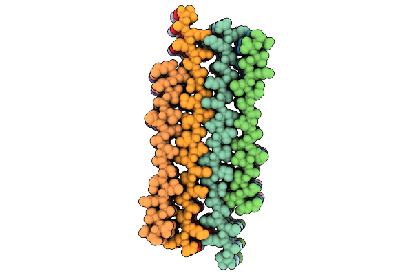
Method: ELECTRON MICROSCOPY Release Date: 2025-10-29 Classification: PROTEIN FIBRIL |
 |
Organism: Homo sapiens
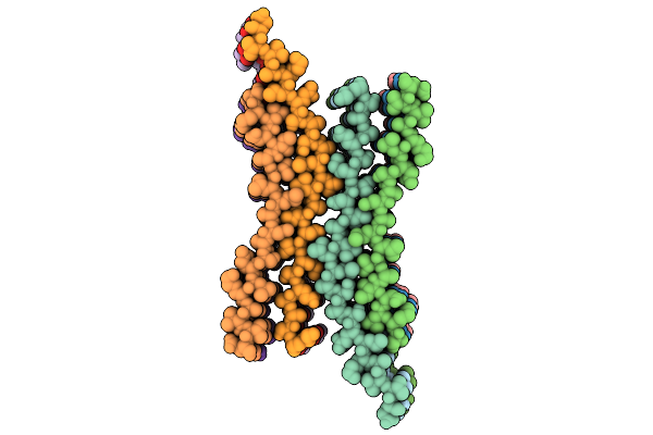
Method: ELECTRON MICROSCOPY Release Date: 2025-10-29 Classification: PROTEIN FIBRIL |
 |
Organism: Saccharomyces cerevisiae s288c
Method: ELECTRON MICROSCOPY Release Date: 2024-04-17 Classification: HYDROLASE |
 |
Organism: Saccharomyces cerevisiae s288c
Method: ELECTRON MICROSCOPY Release Date: 2024-04-17 Classification: HYDROLASE |
 |
Organism: Saccharomyces cerevisiae s288c
Method: ELECTRON MICROSCOPY Release Date: 2022-08-10 Classification: HYDROLASE |
 |
Cryo-Em Structure Of The 20S Alpha 3 Deletion Proteasome Core Particle In Complex With Fub1
Organism: Saccharomyces cerevisiae s288c
Method: ELECTRON MICROSCOPY Release Date: 2022-08-10 Classification: HYDROLASE |
 |
Organism: Severe acute respiratory syndrome coronavirus 2, Homo sapiens
Method: ELECTRON MICROSCOPY Release Date: 2021-12-15 Classification: VIRAL PROTEIN Ligands: NAG |
 |
Organism: Severe acute respiratory syndrome coronavirus 2
Method: ELECTRON MICROSCOPY Release Date: 2021-12-15 Classification: VIRAL PROTEIN Ligands: NAG |
 |
Structural Basis Of Rack7 Phd To Read A Pediatric Glioblastoma-Associated Histone Mutation H3.3G34R
Organism: Homo sapiens
Method: SOLUTION NMR Release Date: 2021-05-26 Classification: TRANSCRIPTION Ligands: ZN |
 |
Crystal Structure Of Sars-Cov-2 Hr1 Motif In Complex With Pan-Covs Inhibitor Ek1
Organism: Severe acute respiratory syndrome coronavirus 2, Synthetic construct
Method: X-RAY DIFFRACTION Resolution:2.28 Å Release Date: 2021-05-19 Classification: VIRAL PROTEIN Ligands: CA |
 |
Organism: Mus musculus
Method: X-RAY DIFFRACTION Resolution:2.05 Å Release Date: 2020-02-05 Classification: TOXIN Ligands: GOL, CL |
 |
Organism: Mus musculus
Method: ELECTRON MICROSCOPY Release Date: 2020-02-05 Classification: TOXIN Ligands: NAG |
 |
Organism: Mus musculus
Method: X-RAY DIFFRACTION Resolution:3.17 Å Release Date: 2020-02-05 Classification: TOXIN |
 |
Organism: Mus musculus
Method: ELECTRON MICROSCOPY Release Date: 2020-02-05 Classification: TOXIN Ligands: NAG |