
Search Count: 941
 |
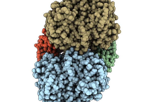
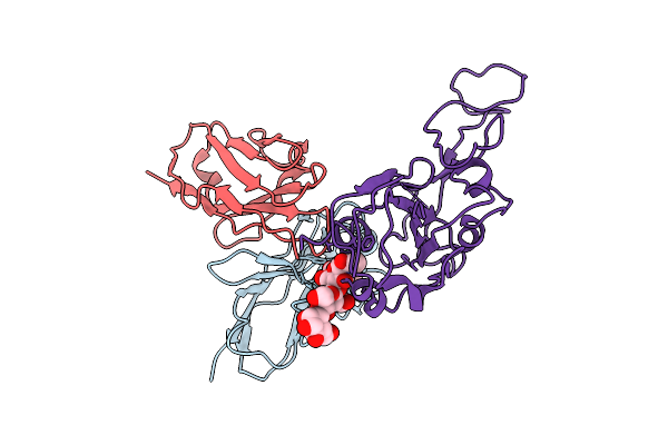
Organism: Escherichia coli
Method: X-RAY DIFFRACTION Release Date: 2026-01-14 Classification: TRANSFERASE Ligands: GOL, MN, AMP, SO4 |
 |
Organism: Escherichia coli
Method: X-RAY DIFFRACTION Release Date: 2026-01-14 Classification: TRANSFERASE Ligands: AMP, SO4 |
 |
Organism: Escherichia coli
Method: X-RAY DIFFRACTION Release Date: 2026-01-14 Classification: LYASE Ligands: NAD, SO4 |
 |
Organism: Mus musculus
Method: X-RAY DIFFRACTION Release Date: 2026-01-14 Classification: LIPID BINDING PROTEIN Ligands: GDP, GOL |
 |
Organism: Mus musculus
Method: X-RAY DIFFRACTION Release Date: 2026-01-14 Classification: LIPID BINDING PROTEIN Ligands: GOL, GSP, MG |
 |
Organism: Mus musculus
Method: X-RAY DIFFRACTION Release Date: 2026-01-14 Classification: LIPID BINDING PROTEIN Ligands: GDP |
 |
Organism: Mus musculus
Method: X-RAY DIFFRACTION Release Date: 2026-01-14 Classification: LIPID BINDING PROTEIN Ligands: GDP |
 |
Organism: Mus musculus
Method: ELECTRON MICROSCOPY Release Date: 2026-01-14 Classification: LIPID BINDING PROTEIN |
 |
Organism: Novosphingopyxis baekryungensis
Method: ELECTRON MICROSCOPY Release Date: 2026-01-14 Classification: CELL INVASION Ligands: MG |
 |
Organism: Bacteriophage sp., Novosphingopyxis baekryungensis
Method: ELECTRON MICROSCOPY Release Date: 2026-01-14 Classification: CELL INVASION |
 |
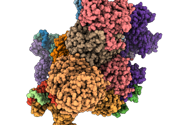
Organism: Severe acute respiratory syndrome coronavirus 2, Homo sapiens
Method: X-RAY DIFFRACTION Release Date: 2026-01-07 Classification: VIRAL PROTEIN/IMMUNE SYSTEM Ligands: NAG |
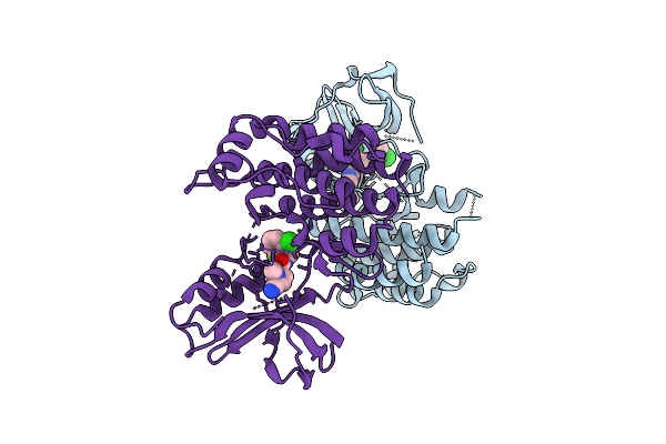 |
Crystal Structure Of An Allosteric Inhibitor Bound To Human Ripk1 Kinase Domain
Organism: Homo sapiens
Method: X-RAY DIFFRACTION Release Date: 2025-12-24 Classification: IMMUNE SYSTEM Ligands: A1IX7, IOD, 1HX |
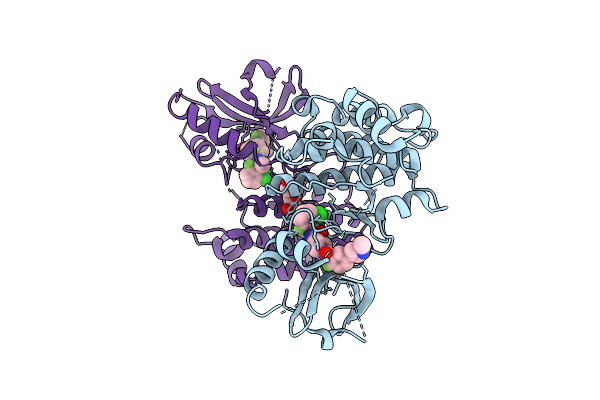 |
Crystal Structure Of An Allosteric Inhibitor Bound To Human Ripk1 Kinase Domain
Organism: Homo sapiens
Method: X-RAY DIFFRACTION Release Date: 2025-12-24 Classification: IMMUNE SYSTEM Ligands: A1IX9, IOD, PEG |
 |
Cryo-Em Structure Of Sars-Cov-2 Ba.5 Spike Protein In Complex With Nab 1C4 (Local Refinement)
Organism: Severe acute respiratory syndrome coronavirus 2, Mus musculus
Method: ELECTRON MICROSCOPY Release Date: 2025-12-10 Classification: VIRAL PROTEIN/IMMUNE SYSTEM |
 |
Cryo-Em Structure Of Sars-Cov-2 Spike Protein In Complex With Three-Nab 8H12, 3E2 And 1C4
Organism: Severe acute respiratory syndrome coronavirus 2, Mus musculus
Method: ELECTRON MICROSCOPY Release Date: 2025-12-10 Classification: VIRAL PROTEIN/IMMUNE SYSTEM |
 |
Cryo-Em Structure Of Sars-Cov-2 Ba.1 Spike Protein In Complex With Three-Nab 8H12, 3E2 And 1C4
Organism: Severe acute respiratory syndrome coronavirus 2, Mus musculus
Method: ELECTRON MICROSCOPY Release Date: 2025-12-10 Classification: VIRAL PROTEIN/IMMUNE SYSTEM |
 |
Cryo-Em Structure Of Sars-Cov-2 Ba.2 Spike Protein In Complex With Triple-Nab 8H12, 3E2 And 1C4 (Local Refinement)
Organism: Severe acute respiratory syndrome coronavirus 2, Mus musculus
Method: ELECTRON MICROSCOPY Release Date: 2025-12-10 Classification: VIRAL PROTEIN/IMMUNE SYSTEM |
 |
Cryo-Em Structure Of Conivaptan-Bound Human Vasopressin V2 Receptor Complex With Fab
Organism: Homo sapiens, Escherichia coli
Method: ELECTRON MICROSCOPY Release Date: 2025-12-10 Classification: MEMBRANE PROTEIN/IMMUNE SYSTEM Ligands: A1ECE |
 |
Cryo-Em Structure Of Tolvaptan-Bound Human Vasopressin V2 Receptor Complex With Fab
Organism: Homo sapiens, Escherichia coli
Method: ELECTRON MICROSCOPY Release Date: 2025-12-10 Classification: MEMBRANE PROTEIN/IMMUNE SYSTEM Ligands: A1ECF |
 |
Cryo-Em Structure Of Adriforant-Bound Histamine Receptor 4 H4R At Inactive State
Organism: Homo sapiens, Escherichia coli
Method: ELECTRON MICROSCOPY Release Date: 2025-12-03 Classification: MEMBRANE PROTEIN/IMMUNE SYSTEM Ligands: A1EBW |