
Search Count: 189
 |
Crystal Structure Of Nodd-Ebd (Effector Binding Domain) In Complex With Hesperetin From Rhizobium Leguminosarum Bv. Vicae 3841
Organism: Rhizobium leguminosarum
Method: X-RAY DIFFRACTION Release Date: 2025-12-24 Classification: TRANSCRIPTION Ligands: GOL, 6JP |
 |
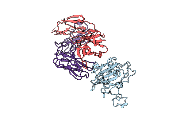
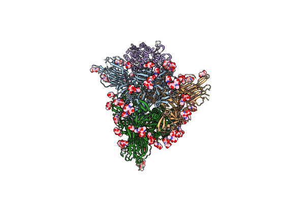
Organism: Severe acute respiratory syndrome coronavirus 2, Homo sapiens
Method: ELECTRON MICROSCOPY Release Date: 2025-12-24 Classification: VIRAL PROTEIN/HYDROLASE |
 |
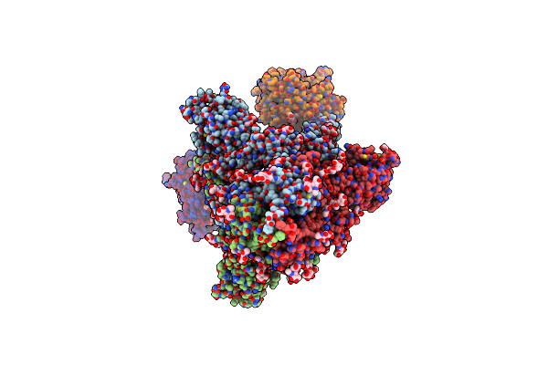
Organism: Homo sapiens, Severe acute respiratory syndrome coronavirus 2
Method: ELECTRON MICROSCOPY Release Date: 2025-12-24 Classification: VIRAL PROTEIN/HYDROLASE |
 |
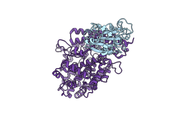
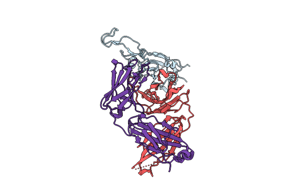
Crystal Structure Of Nodd-Ebd (Effector Binding Domain) From Rhizobium Leguminosarum Bv. Vicae 3841
Organism: Rhizobium leguminosarum
Method: X-RAY DIFFRACTION Release Date: 2025-12-24 Classification: TRANSCRIPTION |
 |
The Local Refined Map Of Sars-Cov-2 Eg.5.1 Variant Spike Protein Complexed With Antibody Xgi-171
Organism: Homo sapiens, Severe acute respiratory syndrome coronavirus 2
Method: ELECTRON MICROSCOPY Release Date: 2025-12-10 Classification: VIRAL PROTEIN/IMMUNE SYSTEM |
 |
The Local Refined Map Of Sars-Cov-2 Eg.5.1 Variant Spike Protein Complexed With Antibody Xgi-183
Organism: Homo sapiens, Severe acute respiratory syndrome coronavirus 2
Method: ELECTRON MICROSCOPY Release Date: 2025-12-10 Classification: VIRAL PROTEIN/IMMUNE SYSTEM |
 |
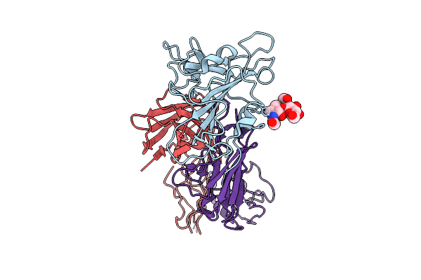
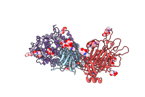
Structure Of Sars-Cov-2 Eg.5.1 Variant Spike Protein Complexed With Antibody Xgi-171
Organism: Homo sapiens, Severe acute respiratory syndrome coronavirus 2
Method: ELECTRON MICROSCOPY Release Date: 2025-12-10 Classification: VIRAL PROTEIN/IMMUNE SYSTEM Ligands: NAG |
 |
The Local Refined Map Of Sars-Cov-2 Eg.5.1 Variant Spike Protein Complexed With Antibody Xgi-198
Organism: Homo sapiens, Severe acute respiratory syndrome coronavirus 2
Method: ELECTRON MICROSCOPY Release Date: 2025-12-10 Classification: VIRAL PROTEIN/IMMUNE SYSTEM |
 |
The Local Refined Map Of Sars-Cov-2 Eg.5.1 Variant Spike Protein Complexed With Antibody Xgi-203
Organism: Severe acute respiratory syndrome coronavirus 2, Homo sapiens
Method: ELECTRON MICROSCOPY Release Date: 2025-12-10 Classification: VIRAL PROTEIN/IMMUNE SYSTEM |
 |
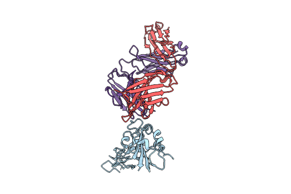
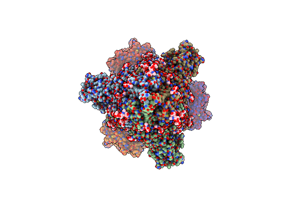
Structure Of Sars-Cov-2 Eg.5.1 Variant Spike Protein Complexed With Antibody Xgi-203
Organism: Homo sapiens, Severe acute respiratory syndrome coronavirus 2
Method: ELECTRON MICROSCOPY Release Date: 2025-12-10 Classification: VIRAL PROTEIN/IMMUNE SYSTEM Ligands: NAG |
 |
Structure Of Sars-Cov-2 Eg.5.1 Variant Spike Protein Complexed With Antibody Xgi-198
Organism: Severe acute respiratory syndrome coronavirus 2, Homo sapiens
Method: ELECTRON MICROSCOPY Release Date: 2025-12-10 Classification: VIRAL PROTEIN/IMMUNE SYSTEM Ligands: NAG |
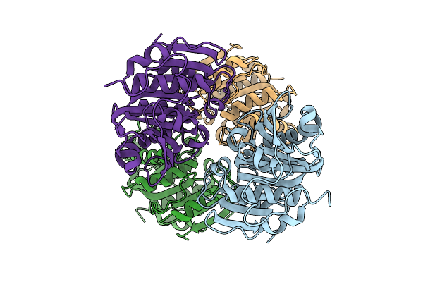 |
Organism: Kibdelosporangium banguiense
Method: X-RAY DIFFRACTION Release Date: 2025-07-30 Classification: HYDROLASE |
 |
 |
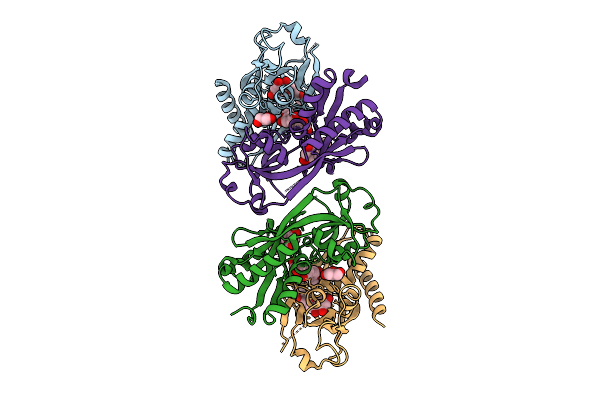
Organism: Shigella sonnei
Method: X-RAY DIFFRACTION Resolution:2.25 Å Release Date: 2025-03-05 Classification: TOXIN Ligands: ACO, PO4 |
 |
Organism: Shigella sonnei
Method: X-RAY DIFFRACTION Resolution:2.75 Å Release Date: 2025-03-05 Classification: TOXIN Ligands: PO4 |
 |
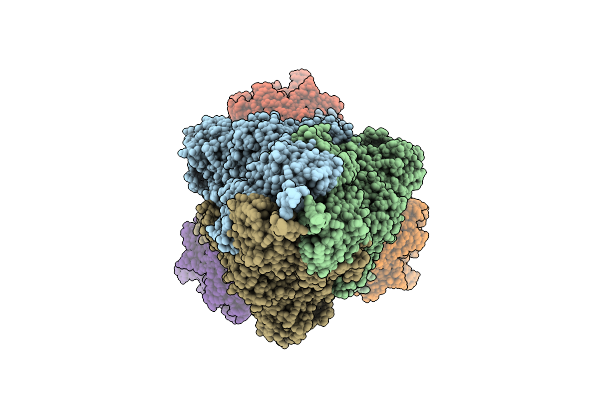
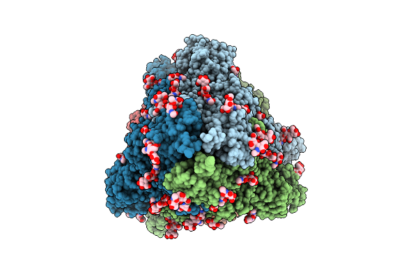
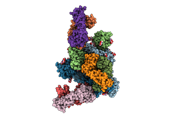
Cryo-Em Structure Of Sars-Cov-2 D614G S With One Ace2 Receptor Binding (Rb1) In Prefusion Conformation
Organism: Severe acute respiratory syndrome coronavirus 2, Homo sapiens
Method: ELECTRON MICROSCOPY Release Date: 2025-02-05 Classification: VIRAL PROTEIN/HYDROLASE Ligands: NAG |
 |
Cryo-Em Structure Of Sars-Cov-2 D614G S With Two Ace2 Receptors Binding (Rb2) In Prefusion Conformation
Organism: Severe acute respiratory syndrome coronavirus 2, Homo sapiens
Method: ELECTRON MICROSCOPY Release Date: 2025-02-05 Classification: VIRAL PROTEIN/HYDROLASE Ligands: NAG |
 |
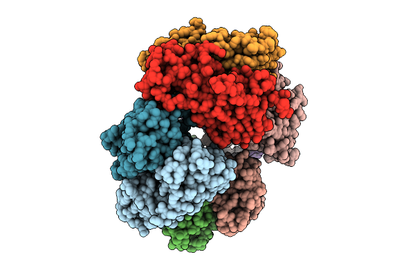
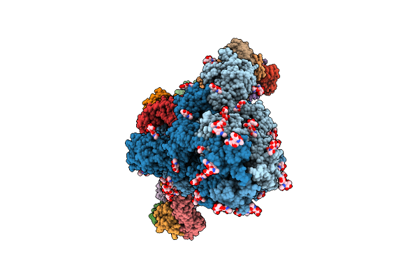
Cryo-Em Structure Of Sars-Cov-2 D614G S With Three Ace2 Receptors Binding (Rb3) In Prefusion Conformation (Focused Refinement Of Ntd-Sd1-Rbd-Ace2)
Organism: Severe acute respiratory syndrome coronavirus 2, Homo sapiens
Method: ELECTRON MICROSCOPY Release Date: 2025-02-05 Classification: VIRAL PROTEIN/HYDROLASE Ligands: NAG |
 |
Cryo-Em Structure Of Sars-Cov-2 D614G S With Three Ace2 Receptors Binding (Rb3) In Prefusion Conformation
Organism: Severe acute respiratory syndrome coronavirus 2, Homo sapiens
Method: ELECTRON MICROSCOPY Release Date: 2025-02-05 Classification: VIRAL PROTEIN/HYDROLASE Ligands: NAG |
 |
Cryo-Em Structure Of Sars-Cov-2 S Trimer In The Early Fusion Intermediate Conformation (E-Fic) (Focused Refinement Of Ntd-Sd1-Rbd-Ace2)
Organism: Homo sapiens, Severe acute respiratory syndrome coronavirus 2
Method: ELECTRON MICROSCOPY Release Date: 2025-02-05 Classification: VIRAL PROTEIN Ligands: NAG |