
Search Count: 26
 |
Organism: Synthetic construct
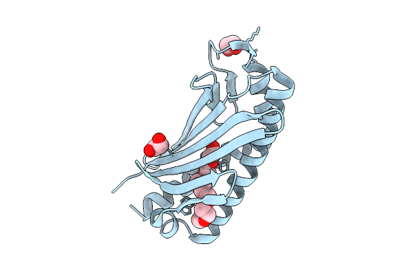
Method: X-RAY DIFFRACTION Resolution:1.60 Å Release Date: 2025-08-13 Classification: DE NOVO PROTEIN Ligands: EDO, P15 |
 |
Organism: Synthetic construct
Method: X-RAY DIFFRACTION Resolution:1.85 Å Release Date: 2025-04-02 Classification: OXIDOREDUCTASE Ligands: ZN |
 |
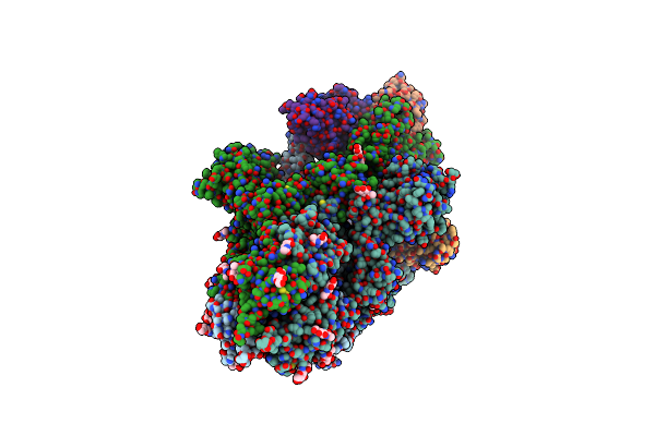
Organism: Severe acute respiratory syndrome coronavirus 2, Homo sapiens
Method: ELECTRON MICROSCOPY Release Date: 2022-11-30 Classification: VIRAL PROTEIN |
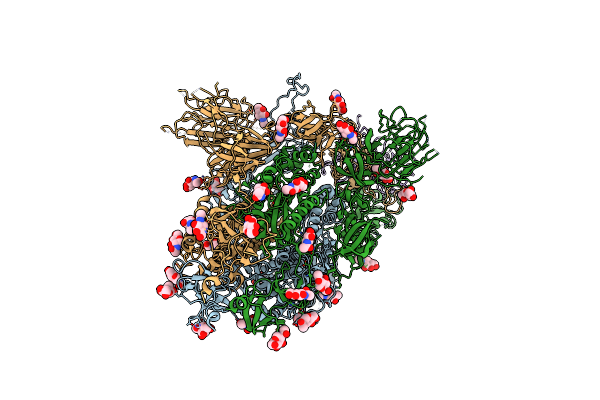 |
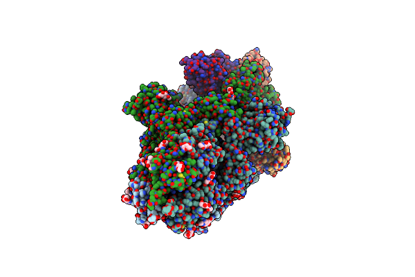
Organism: Severe acute respiratory syndrome coronavirus 2, Homo sapiens
Method: ELECTRON MICROSCOPY Release Date: 2022-11-30 Classification: VIRAL PROTEIN Ligands: NAG |
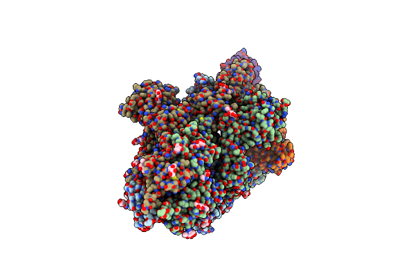 |
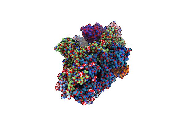
Organism: Severe acute respiratory syndrome coronavirus 2, Homo sapiens
Method: ELECTRON MICROSCOPY Release Date: 2022-11-30 Classification: VIRAL PROTEIN Ligands: NAG |
 |
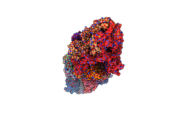
Organism: Severe acute respiratory syndrome coronavirus 2, Homo sapiens
Method: ELECTRON MICROSCOPY Release Date: 2022-11-30 Classification: VIRAL PROTEIN Ligands: NAG |
 |
Organism: Severe acute respiratory syndrome coronavirus 2, Homo sapiens
Method: ELECTRON MICROSCOPY Release Date: 2022-11-30 Classification: VIRAL PROTEIN Ligands: NAG |
 |
Organism: Severe acute respiratory syndrome coronavirus 2, Homo sapiens
Method: ELECTRON MICROSCOPY Release Date: 2022-11-30 Classification: VIRAL PROTEIN Ligands: NAG |
 |
Organism: Severe acute respiratory syndrome coronavirus 2, Homo sapiens
Method: ELECTRON MICROSCOPY Release Date: 2022-11-30 Classification: VIRAL PROTEIN |
 |
Crystal Structure Of An Esterase From The Bacterial Hormone-Sensitive Lipase (Hsl) Family
Organism: Environmental samples
Method: X-RAY DIFFRACTION Resolution:2.00 Å Release Date: 2015-03-25 Classification: HYDROLASE Ligands: PMS |
 |
Organism: Uncultured bacterium
Method: X-RAY DIFFRACTION Resolution:2.05 Å Release Date: 2014-06-04 Classification: HYDROLASE |
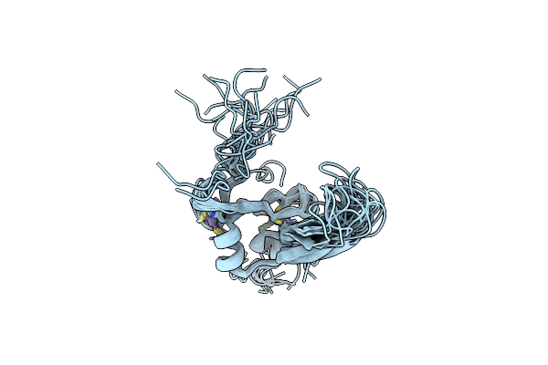 |
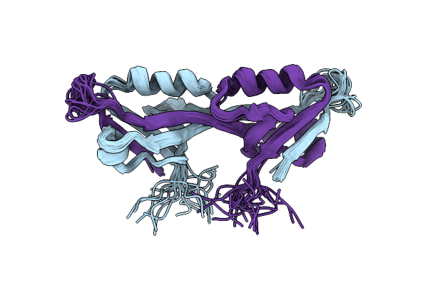
Organism: Homo sapiens
Method: SOLUTION NMR Release Date: 2012-10-24 Classification: METAL BINDING PROTEIN Ligands: ZN |
 |
Organism: Homo sapiens
Method: SOLUTION NMR Release Date: 2012-08-08 Classification: RNA BINDING PROTEIN |
 |
Organism: Homo sapiens
Method: SOLUTION NMR Release Date: 2011-03-02 Classification: SIGNALING PROTEIN |
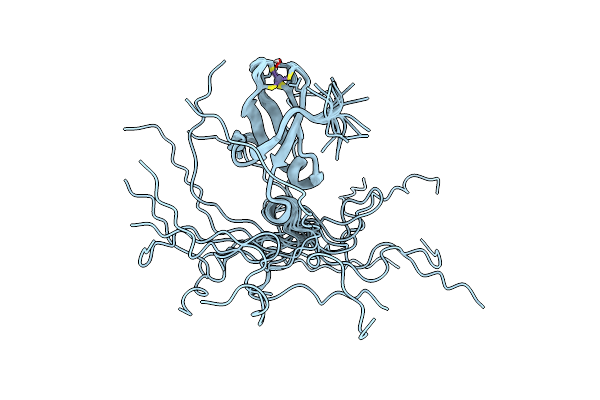 |
Organism: Homo sapiens
Method: SOLUTION NMR Release Date: 2008-03-25 Classification: METAL BINDING PROTEIN Ligands: ZN |
 |
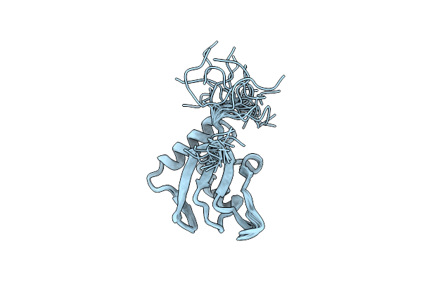
Solution Structure Of The Second Pdz Domain From Human Zonula Occludens-1: A Dimeric Form With 3D Domain Swapping
|
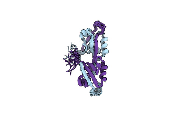 |
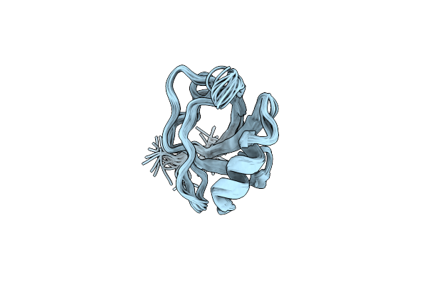
Solution Structure And Binding Property Of The Domain-Swapped Dimer Of Zo2Pdz2
|
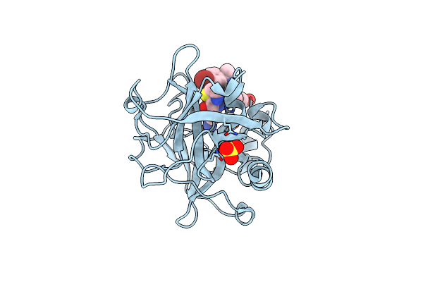 |
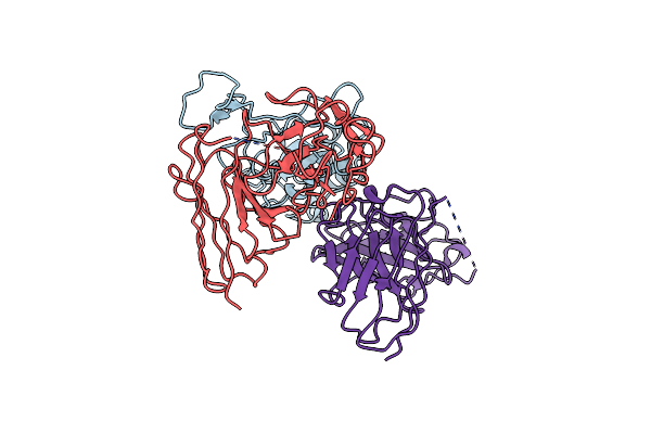
Crystal Structure Of The Catalytic Domain Of Coagulation Factor Xi In Complex With A Peptidomimetic Inhibitor
Organism: Homo sapiens
Method: X-RAY DIFFRACTION Resolution:2.25 Å Release Date: 2006-05-23 Classification: HYDROLASE Ligands: SO4, 339 |
 |
Organism: Homo sapiens
Method: X-RAY DIFFRACTION Resolution:2.05 Å Release Date: 2006-05-09 Classification: HYDROLASE Ligands: 624 |
 |
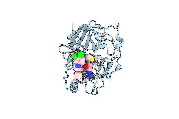
Crystal Structure Of The Catalytic Domain Of Coagulation Factor Xi In Complex With 2-(5-Benzylamino-2-Methylsulfanyl-6-Oxo-6H-Pyrimidin-1-Yl)-N-[4-Guanidino-1-(Thiazole-2-Carbonyl)-Butyl]-Acetamide
Organism: Homo sapiens
Method: X-RAY DIFFRACTION Resolution:2.05 Å Release Date: 2006-05-09 Classification: HYDROLASE Ligands: SO4, BCT, 632 |