
Search Count: 17
 |
Organism: Synthetic construct
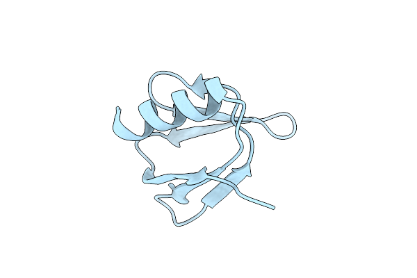
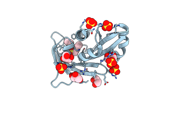
Method: X-RAY DIFFRACTION Resolution:1.21 Å Release Date: 2023-03-22 Classification: DE NOVO PROTEIN |
 |
Organism: Synthetic construct
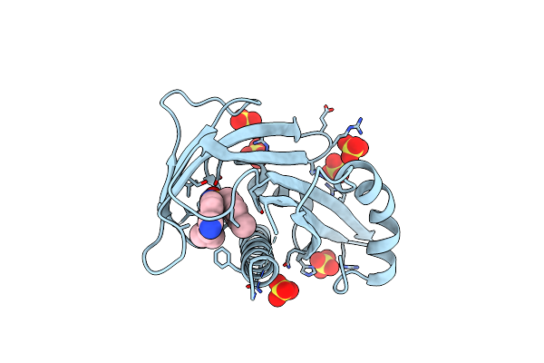
Method: X-RAY DIFFRACTION Resolution:1.50 Å Release Date: 2023-03-22 Classification: DE NOVO PROTEIN |
 |
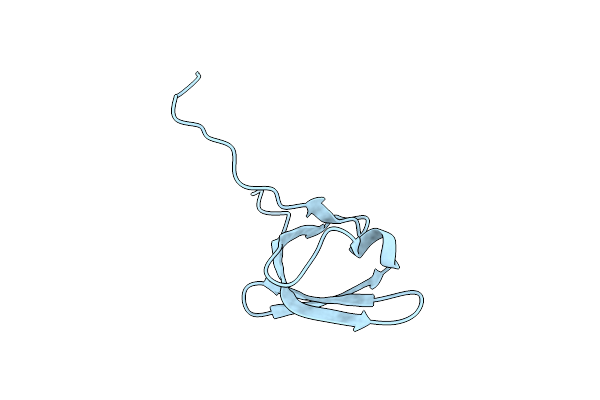
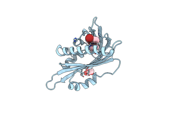
Nmr Solution Structure Of The De Novo Designed Small Beta-Barrel Protein 29_Bp_Sh3
Organism: Synthetic construct
Method: SOLUTION NMR Release Date: 2023-03-22 Classification: DE NOVO PROTEIN |
 |
Nmr Solution Structure Of The De Novo Designed Small Beta-Barrel Protein 33_Bp_Sh3
Organism: Synthetic construct
Method: SOLUTION NMR Release Date: 2023-03-22 Classification: DE NOVO PROTEIN |
 |
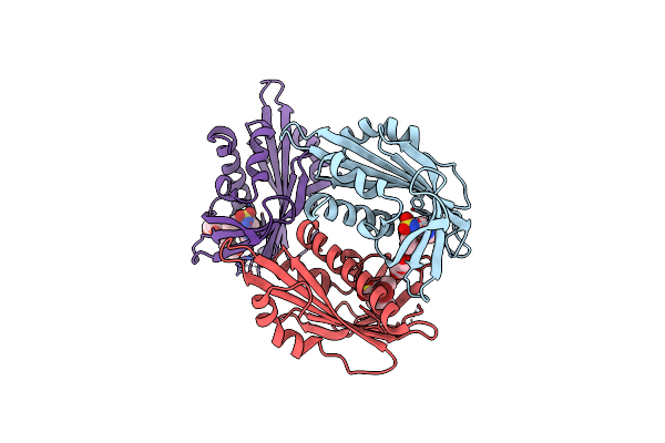
Organism: Arabidopsis thaliana
Method: X-RAY DIFFRACTION Resolution:1.93 Å Release Date: 2017-11-29 Classification: HORMONE RECEPTOR Ligands: SO4, 8KP, GOL |
 |
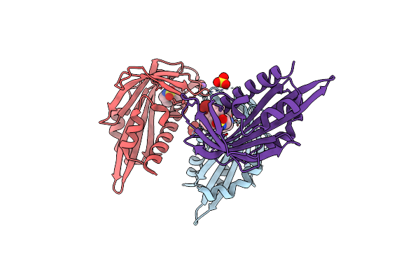
Organism: Arabidopsis thaliana
Method: X-RAY DIFFRACTION Resolution:1.63 Å Release Date: 2017-11-22 Classification: HORMONE RECEPTOR Ligands: SO4, 8KM |
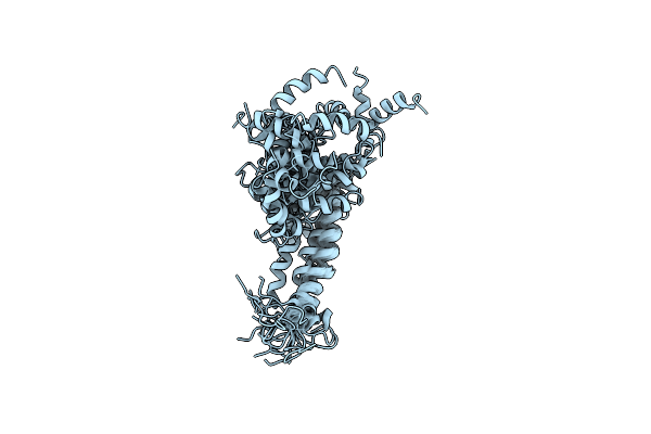 |
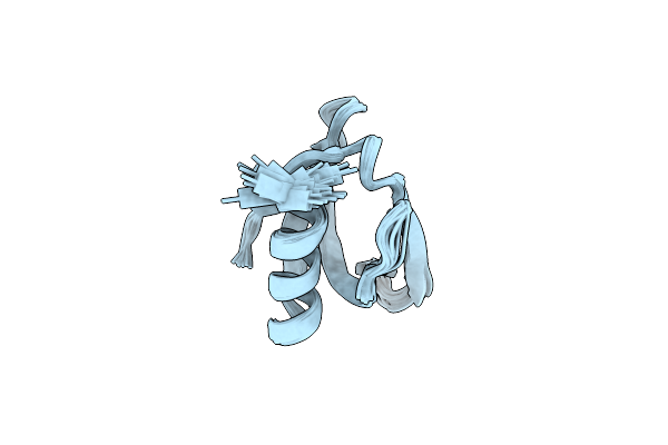
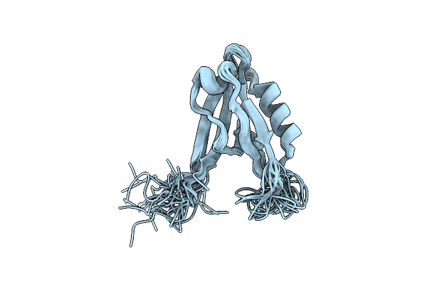
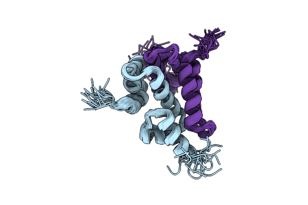
Structure Of The C-Terminal Transmembrane Domain Of Scavenger Receptor Bi (Sr-Bi)
Organism: Mus musculus
Method: SOLUTION NMR Release Date: 2017-03-08 Classification: SIGNALING PROTEIN |
 |
 |
Organism: Arabidopsis thaliana
Method: X-RAY DIFFRACTION Resolution:1.89 Å Release Date: 2010-08-18 Classification: HORMONE RECEPTOR Ligands: PYV, PEG, GOL |
 |
Organism: Arabidopsis thaliana
Method: X-RAY DIFFRACTION Resolution:1.95 Å Release Date: 2010-08-18 Classification: HORMONE RECEPTOR Ligands: PYV, P2M, GOL |
 |
Organism: Arabidopsis thaliana
Method: X-RAY DIFFRACTION Resolution:2.47 Å Release Date: 2010-08-18 Classification: HORMONE RECEPTOR Ligands: PYV, SO4, NA, P2M, CL |
 |
Organism: Arabidopsis thaliana
Method: X-RAY DIFFRACTION Resolution:2.40 Å Release Date: 2009-12-08 Classification: SIGNALING PROTEIN |
 |
Organism: Arabidopsis thaliana
Method: X-RAY DIFFRACTION Resolution:1.85 Å Release Date: 2009-12-08 Classification: SIGNALING PROTEIN Ligands: BU2 |
 |
Organism: Arabidopsis thaliana
Method: X-RAY DIFFRACTION Resolution:1.95 Å Release Date: 2009-12-08 Classification: SIGNALING PROTEIN Ligands: A8S |
 |
Organism: Arabidopsis thaliana
Method: X-RAY DIFFRACTION Resolution:1.95 Å Release Date: 2009-12-08 Classification: SIGNALING PROTEIN Ligands: A8S, MG |
 |
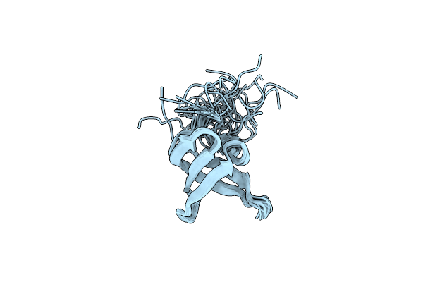
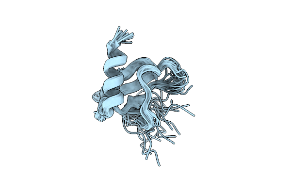
Organism: Homo sapiens
Method: SOLUTION NMR Release Date: 2009-11-10 Classification: SIGNALING PROTEIN |
 |