
Search Count: 18
 |
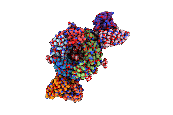
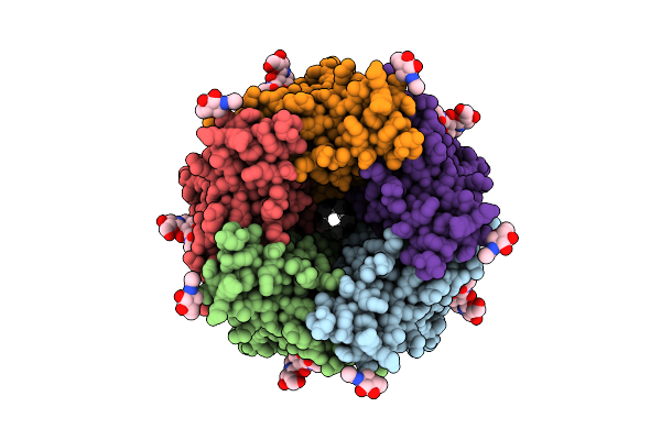
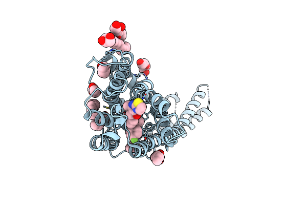
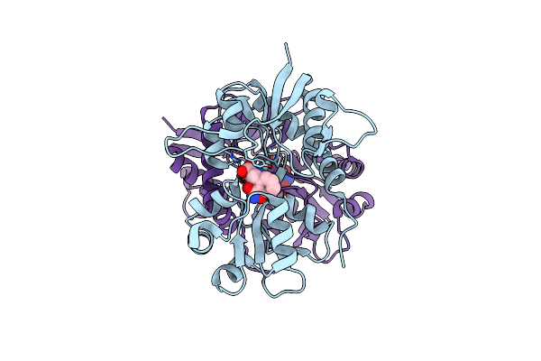
Human Gabaa Receptor Alpha1-Beta2-Gamma2 Subtype In Complex With Gaba Plus Methaqualone
Organism: Homo sapiens, Mus musculus
Method: ELECTRON MICROSCOPY Release Date: 2024-06-26 Classification: MEMBRANE PROTEIN Ligands: ABU, A1ADG, PTY, Q3G |
 |
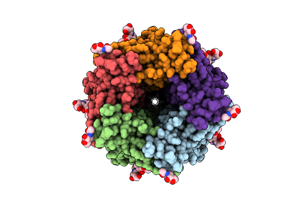
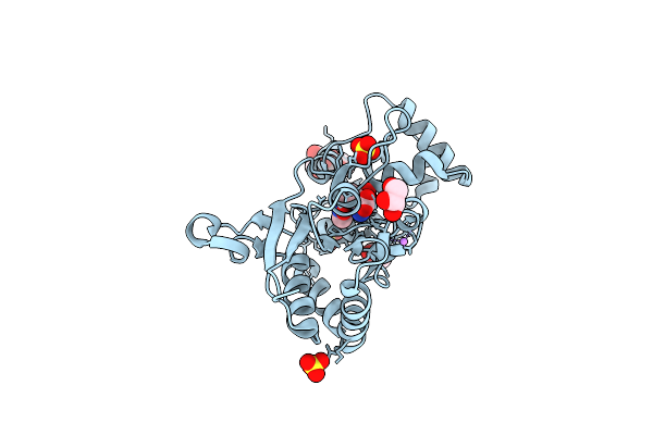
Human Gabaa Receptor Alpha1-Beta2-Gamma2 Subtype In Complex With Gaba Plus Pptq
Organism: Homo sapiens, Mus musculus
Method: ELECTRON MICROSCOPY Release Date: 2024-06-26 Classification: MEMBRANE PROTEIN Ligands: ABU, A1ADN, PTY, POV, Q3G |
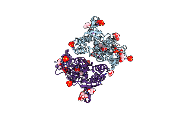 |
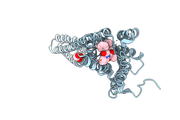
Structure Of Glua2 Ligand-Binding Domain (S1S2J) In Complex With The Agonist (S)-2-Amino-3-(1-Ethyl-4-Hydroxy-1H-1,2,3-Triazol-5-Yl)Propanoic Acid At 1.4 A Resolution
Organism: Rattus norvegicus
Method: X-RAY DIFFRACTION Resolution:1.40 Å Release Date: 2019-04-17 Classification: MEMBRANE PROTEIN Ligands: SO4, HJ8, GOL, PGE, CL, CIT, LI, PEG |
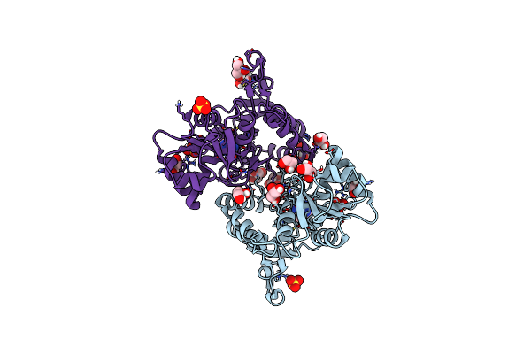 |
Structure Of Glua2 Ligand-Binding Domain (S1S2J) In Complex With The Agonist (S)-2-Amino-3-(2-Methyl-5-Hydroxy-2H-1,2,3-Triazol-4-Yl)Propanoic Acid At 1.55 A Resolution
Organism: Rattus norvegicus
Method: X-RAY DIFFRACTION Resolution:1.55 Å Release Date: 2019-04-17 Classification: MEMBRANE PROTEIN Ligands: HJH, SO4, GOL, CL, LI |
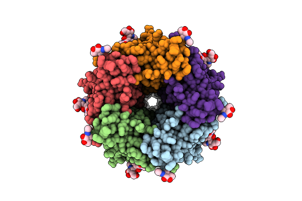 |
Organism: Mus musculus
Method: ELECTRON MICROSCOPY Release Date: 2018-11-07 Classification: MEMBRANE PROTEIN Ligands: NAG, SRO |
 |
Organism: Mus musculus
Method: ELECTRON MICROSCOPY Release Date: 2018-11-07 Classification: MEMBRANE PROTEIN Ligands: NAG, SRO |
 |
Organism: Mus musculus
Method: ELECTRON MICROSCOPY Release Date: 2018-11-07 Classification: MEMBRANE PROTEIN Ligands: NAG, SRO |
 |
Organism: Mus musculus
Method: ELECTRON MICROSCOPY Release Date: 2018-11-07 Classification: MEMBRANE PROTEIN Ligands: TKT |
 |
Organism: Homo sapiens, Escherichia coli
Method: X-RAY DIFFRACTION Resolution:3.00 Å Release Date: 2018-02-14 Classification: MEMBRANE PROTEIN Ligands: ERM, OLC |
 |
Organism: Homo sapiens, Escherichia coli
Method: X-RAY DIFFRACTION Resolution:2.70 Å Release Date: 2018-02-14 Classification: MEMBRANE PROTEIN Ligands: E2J, OLC, PEG, OLA |
 |
Crystal Structure Of The Glua2 Ligand-Binding Domain (S1S2J) In Complex With (S)-2-Amino-3-(5-(2-(3-(Aminomethyl)Benzyl)-2H-Tetrazol-5-Yl)-3-Hydroxyisoxazol-4-Yl)Propanoic Acid At Resolution 1.55 A Resolution
Organism: Rattus norvegicus
Method: X-RAY DIFFRACTION Resolution:1.55 Å Release Date: 2016-03-02 Classification: MEMBRANE PROTEIN Ligands: SO4, 5XP, GOL, ACT, EDO, LI |
 |
Crystal Structure Of The Glua2 Ligand-Binding Domain (S1S2J) In Complex With (S)-2-Amino-3-(5-(2-(3-Methylbenzyl)-2H-Tetrazol-5-Yl)-3-Hydroxyisoxazol-4-Yl)Propanoic Acid At 1.6 A Resolution
Organism: Rattus norvegicus
Method: X-RAY DIFFRACTION Resolution:1.60 Å Release Date: 2016-03-02 Classification: MEMBRANE PROTEIN Ligands: SO4, GOL, ACT, 5XO, EDO |
 |
Crystal Structure Of The Glua2 Ligand-Binding Domain (S1S2J) In Complex With (S)-2-Amino-3-(5-(2-(3-Chlorobenzyl)-2H-Tetrazol-5-Yl)-3-Hydroxyisoxazol-4-Yl)Propanoic Acid At 2.3 A Resolution
Organism: Rattus norvegicus
Method: X-RAY DIFFRACTION Resolution:2.30 Å Release Date: 2016-03-02 Classification: MEMBRANE PROTEIN Ligands: 5XN, SO4, GOL, CL, EDO, PG4 |
 |
Crystal Structure Of The Kainate Receptor Gluk3 Ligand-Binding Domain In Complex With The Agonist Za302
Organism: Rattus norvegicus
Method: X-RAY DIFFRACTION Resolution:2.65 Å Release Date: 2013-03-06 Classification: MEMBRANE PROTEIN Ligands: 3ZA, K, CL, PO4 |
 |
Crystal Structure Of The Glua2 Ligand-Binding Domain (S1S2J) In Complex With The Agonist Za302 At 1.24A Resolution
Organism: Rattus norvegicus
Method: X-RAY DIFFRACTION Resolution:1.24 Å Release Date: 2013-03-06 Classification: MEMBRANE PROTEIN Ligands: 3ZA, SO4, LI, GOL |
 |
Crystal Structure Of Ls-Achbp Complexed With Carbamoylcholine Analogue 3-(Dimethylamino)Butyl Dimethylcarbamate (Dmabc)
Organism: Lymnaea stagnalis
Method: X-RAY DIFFRACTION Resolution:2.48 Å Release Date: 2013-02-20 Classification: ACETYLCHOLINE-BINDING PROTEIN Ligands: XRX, SO4, NAG, PEG, 1PE |
 |
Crystal Structure Of Ls-Achbp Complexed With Carbamoylcholine Analogue N,N-Dimethyl-4-(1-Methyl-1H-Imidazol-2-Yloxy)Butan-2-Amine
Organism: Lymnaea stagnalis
Method: X-RAY DIFFRACTION Resolution:2.20 Å Release Date: 2013-02-20 Classification: ACETYLCHOLINE-BINDING PROTEIN Ligands: XRS, SO4, NAG |
 |
Crystal Structure Of The Ligand-Binding Core Of Glur5 In Complex With The Agonist 4-Ahcp
Organism: Rattus norvegicus
Method: X-RAY DIFFRACTION Resolution:2.20 Å Release Date: 2009-07-21 Classification: MEMBRANE PROTEIN Ligands: CL, IBC |