
Search Count: 24
 |
Organism: Homo sapiens
Method: X-RAY DIFFRACTION Resolution:1.90 Å Release Date: 2017-12-20 Classification: METAL BINDING PROTEIN Ligands: FES |
 |
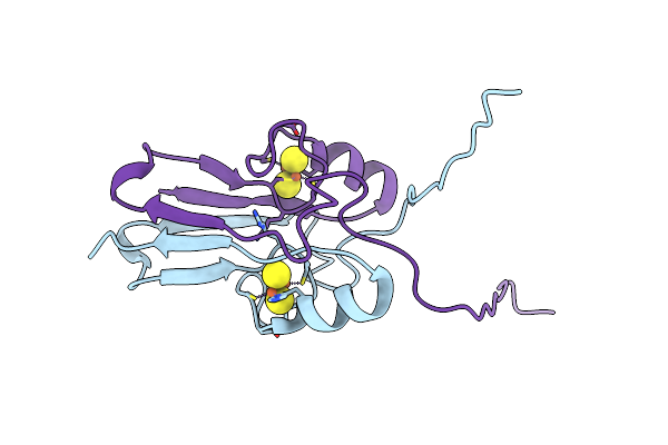
The 1.55A Crystal Structure Of Naf1 (Miner1): The Redox-Active 2Fe-2S Protein
Organism: Homo sapiens
Method: X-RAY DIFFRACTION Resolution:1.65 Å Release Date: 2014-07-02 Classification: METAL BINDING PROTEIN Ligands: FES |
 |
Organism: Homo sapiens
Method: X-RAY DIFFRACTION Resolution:1.58 Å Release Date: 2014-07-02 Classification: METAL BINDING PROTEIN Ligands: FES |
 |
The Crystal Structure Of A Human Mitoneet Mutant With An Ala Inserted Between Asp 67 And Lys 68
Organism: Homo sapiens
Method: X-RAY DIFFRACTION Resolution:1.19 Å Release Date: 2012-12-26 Classification: METAL BINDING PROTEIN Ligands: FES |
 |
The Crystal Structure Of A Human Mitoneet Mutant With Asp 67 Replaced By A Gly
Organism: Homo sapiens
Method: X-RAY DIFFRACTION Resolution:2.40 Å Release Date: 2012-12-26 Classification: METAL BINDING PROTEIN Ligands: FES |
 |
The Crystal Structure Of A Human Mitoneet Mutant With Met 62 Replaced By A Gly
Organism: Homo sapiens
Method: X-RAY DIFFRACTION Resolution:1.55 Å Release Date: 2012-12-26 Classification: METAL BINDING PROTEIN Ligands: FES |
 |
The Crystal Structure Of A Human Mitoneet Double Mutant In Which Gly 66 Are Asp 67 Are Both Replaced With Ala Residues
Organism: Homo sapiens
Method: X-RAY DIFFRACTION Resolution:1.35 Å Release Date: 2012-12-26 Classification: METAL BINDING PROTEIN Ligands: FES |
 |
Organism: Arabidopsis thaliana
Method: X-RAY DIFFRACTION Resolution:1.75 Å Release Date: 2012-05-23 Classification: METAL BINDING PROTEIN Ligands: FES, ZN |
 |
Organism: Arabidopsis thaliana
Method: X-RAY DIFFRACTION Resolution:1.14 Å Release Date: 2012-05-23 Classification: METAL BINDING PROTEIN Ligands: FES |
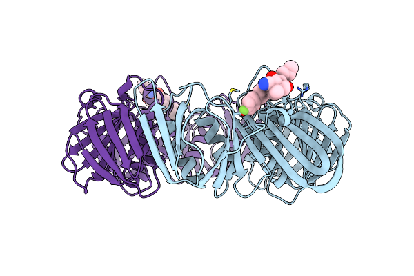 |
Structure Of E. Coli Poliiibeta With (Z)-5-(1-((4'-Fluorobiphenyl-4-Yl)Methoxyimino)Butyl)-2,2-Dimethyl-4,6-Dioxocyclohexanecarbonitrile
Organism: Escherichia coli
Method: X-RAY DIFFRACTION Resolution:1.90 Å Release Date: 2011-06-08 Classification: TRANSFERASE/TRANSFERASE INHIBITOR Ligands: 743 |
 |
Organism: Homo sapiens
Method: X-RAY DIFFRACTION Resolution:1.70 Å Release Date: 2011-06-01 Classification: METAL BINDING PROTEIN Ligands: FES |
 |
Organism: Mastigocladus laminosus
Method: X-RAY DIFFRACTION Resolution:2.30 Å Release Date: 2011-02-09 Classification: ELECTRON TRANSPORT Ligands: FES |
 |
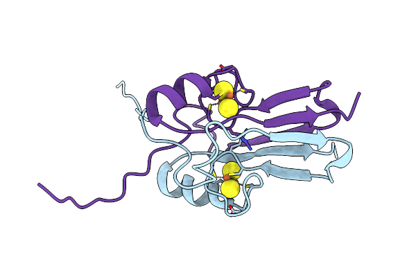
Crystal Structure Of Miner1: The Redox-Active 2Fe-2S Protein Causative In Wolfram Syndrome 2
Organism: Homo sapiens
Method: X-RAY DIFFRACTION Resolution:2.10 Å Release Date: 2009-08-18 Classification: METAL BINDING PROTEIN Ligands: FES |
 |
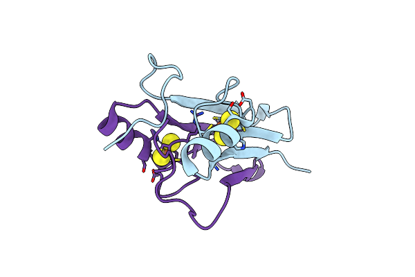
The Novel 2Fe-2S Outer Mitochondrial Protein Mitoneet Displays Conformational Flexibility In Its N-Terminal Cytoplasmic Tethering Domain
Organism: Homo sapiens
Method: X-RAY DIFFRACTION Resolution:1.40 Å Release Date: 2009-07-07 Classification: METAL BINDING PROTEIN Ligands: FES |
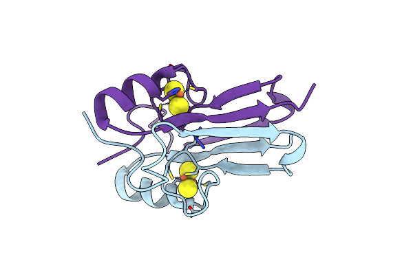 |
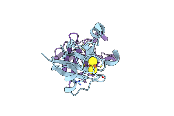
Mitoneet Is A Uniquely Folded 2Fe-2S Outer Mitochondrial Membrane Protein Stabilized By Pioglitazone
Organism: Homo sapiens
Method: X-RAY DIFFRACTION Resolution:1.50 Å Release Date: 2007-08-21 Classification: METAL BINDING PROTEIN Ligands: FES |
 |
Crystal Structure Of Rii Alpha Dimerization/Docking Domain Of Pka Bound To The D-Akap2 Peptide
Organism: Rattus norvegicus
Method: X-RAY DIFFRACTION Resolution:1.60 Å Release Date: 2006-11-21 Classification: TRANSFERASE Ligands: GOL |
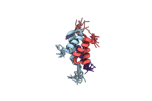 |
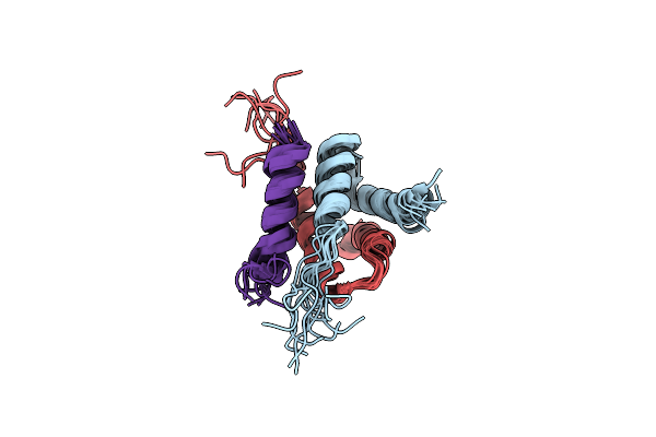
Docking And Dimerization Domain (D/D) Of The Type Ii-Alpha Regulatory Subunity Of Protein Kinase A (Pka) In Complex With A Peptide From An A-Kinase Anchoring Protein
Organism: Rattus norvegicus
Method: SOLUTION NMR Release Date: 2006-08-29 Classification: TRANSFERASE |
 |
Docking And Dimerization Domain (D/D) Of The Regulatory Subunit Of The Type Ii-Alpha Camp-Dependent Protein Kinase A Associated With A Peptide Derived From An A-Kinase Anchoring Protein (Akap)
Organism: Rattus norvegicus
Method: SOLUTION NMR Release Date: 2006-08-29 Classification: TRANSFERASE |
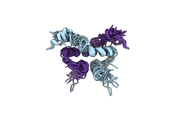 |
Solution Structure Of The Docking And Dimerization Domain Of The Type I Alpha Regulatory Subunit Of Protein Kinase A (Rialpha D/D)
|
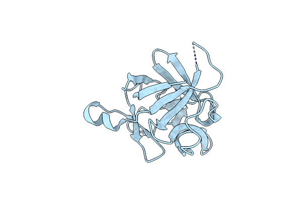 |
Organism: Homo sapiens
Method: X-RAY DIFFRACTION Resolution:1.54 Å Release Date: 2003-02-04 Classification: IMMUNE SYSTEM |